Acute Kidney Injury (AKI) Nursing Care Plan
AKI nursing care plan: fluid balance, electrolyte monitoring, BUN/creatinine trending, and a printable PDF. Built by nurses for nurses.
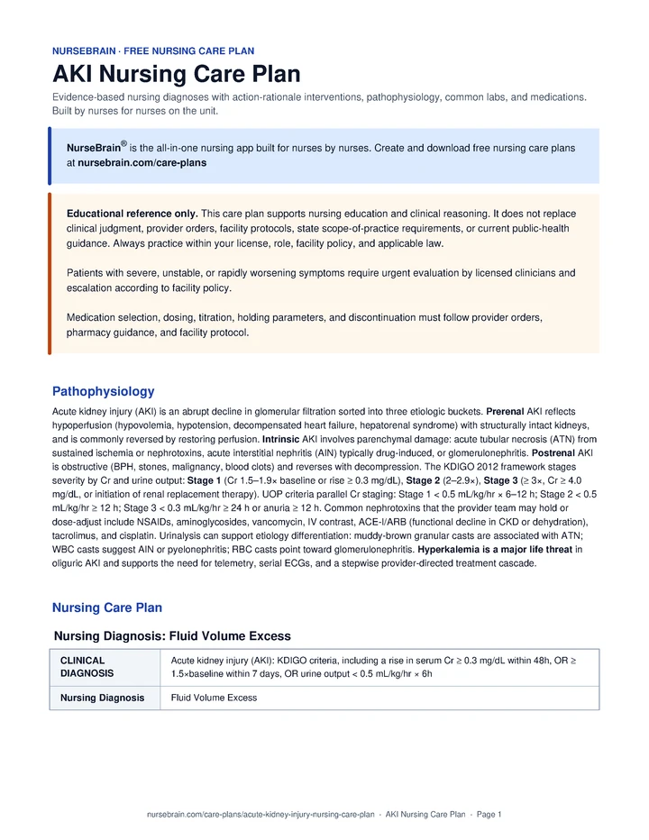
Nursing Care Plan
Nursing Diagnosis 1: Fluid Volume Excess
Fluid Volume Excess related to Acute kidney injury (AKI): KDIGO criteria, including a rise in serum Cr ≥ 0.3 mg/dL within 48h, OR ≥ 1.5×baseline within 7 days, OR urine output < 0.5 mL/kg/hr × 6h as evidenced by Oliguria with UOP < 0.5 mL/kg/hr; Weight gain > 2 kg in 48 hours; Bilateral 2+ pitting lower-extremity edema; Jugular venous distention with HOB at 30–45°; Bibasilar crackles on auscultation.
Interventions
- Maintain strict hourly intake and output via indwelling catheter; calculate and document net 24-hour balance per facility protocol.
- Obtain daily weights at the same time, on the same scale, in the same garments when clinical state allows.
- Auscultate breath sounds in all lung fields at intervals matched to clinical acuity and facility protocol; document new or worsening crackles.
- Assess peripheral edema location, pitting grade (1–4+), and symmetry each shift.
- Monitor BUN, Cr, electrolytes (K+, Mg2+, phosphorus), and bicarbonate per provider order.
- Assess for JVD with head of bed at 30–45° each shift and document findings.
- Implement prescribed fluid restriction (commonly 1–1.5 L/day in oliguric AKI per provider order); coordinate with dietary and the provider team.
- Administer prescribed loop diuretics as ordered for documented volume overload; per KDIGO 2012, diuretics are not recommended for AKI prevention or treatment.
- Position the patient in semi- to high-Fowler’s during periods of dyspnea or crackles per provider order and patient tolerance.
- Coordinate with the renal dietitian for a low-sodium and renal-appropriate diet per provider order.
- Administer prescribed phosphate binders with meals per provider order when hyperphosphatemia is present.
- Teach the patient and family the rationale for fluid restriction and how to track intake using a marked container.
- Educate on reading food labels for sodium and potassium content; reinforce the prescribed sodium target and high-K+ foods to limit per the renal diet (bananas, oranges, tomatoes, potatoes).
- Teach daily home weights post-discharge: same time, same scale, same garments; contact the provider for a gain of more than 2 lb (1 kg) overnight per the discharge plan.
- Notify the provider for UOP < 30 mL/hr for 2 consecutive hours, weight gain > 2 kg in 48 h, or new SpO2 drop below ordered parameters.
- Coordinate with nephrology for RRT (CRRT or IHD) evaluation when AEIOU criteria are met per provider direction.
Outcome: Urine output is monitored and reported within ordered parameters; Weight change is documented and reported per provider order; Peripheral edema is monitored and changes are reported promptly.
Nursing Diagnosis 2: Spiritual Distress
Electrolyte Imbalance related to Acute kidney injury (AKI): KDIGO criteria, including a rise in serum Cr ≥ 0.3 mg/dL within 48h, OR ≥ 1.5×baseline within 7 days, OR urine output < 0.5 mL/kg/hr × 6h as evidenced by Serum K+ ≥ 5.5 mEq/L (hyperkalemia); Peaked T-waves or widening QRS on ECG; Metabolic acidosis (HCO3− < 22 mEq/L); Hyperphosphatemia (phos > 4.5 mg/dL); Hypocalcemia (ionized Ca < 4.6 mg/dL).
Interventions
- Monitor serum K+, phosphorus, ionized calcium, Mg2+, and bicarbonate at least daily and more frequently per provider order if abnormal or post-treatment.
- Place the patient on continuous telemetry per facility protocol; obtain 12-lead ECG for any K+ ≥ 6.0 mEq/L or new arrhythmia per order.
- Assess for clinical signs of hyperkalemia: muscle weakness, paresthesias, palpitations, hypotonia.
- Review the medication record each shift for nephrotoxins (NSAIDs, aminoglycosides, IV contrast) and K+-elevating drugs (ACE-I, ARB, spironolactone, KCl, TMP-SMX); communicate with the provider and pharmacy.
- Assess for signs of hypocalcemia: Chvostek’s, Trousseau’s, perioral numbness, tetany.
- For K+ ≥ 6.0 with ECG changes, administer IV calcium gluconate 1–2 g over 2–3 min per provider order and facility protocol; repeat ECG in 5–10 min.
- Administer insulin 10 units IV with 25–50 g dextrose per provider order to shift K+ intracellularly; recheck glucose Q1h × 6 and K+ at 1 h per facility protocol.
- Administer nebulized albuterol 10–20 mg as an adjunct shift therapy with insulin per provider order.
- Administer prescribed K+ binder (patiromer, Lokelma, or Kayexalate) as ordered for sustained K+ reduction.
- Administer IV sodium bicarbonate per provider order in metabolic acidosis when ordered (commonly considered with pH < 7.20 or symptomatic acidosis).
- Administer K+, Mg2+, and phosphorus replacement per provider order and facility protocol when low; avoid empiric replacement.
- Teach the patient and family to limit high-potassium foods during AKI (bananas, oranges, tomatoes, potatoes, melons, salt substitutes) per the renal diet.
- Educate on avoiding OTC NSAIDs (ibuprofen, naproxen) and nephrotoxic supplements during and after recovery per the discharge plan.
- Teach recognition of hyperkalemia symptoms: muscle weakness, palpitations, tingling around the mouth; instruct to seek emergency care if symptoms develop.
- Notify the provider promptly for K+ ≥ 6.0 mEq/L, any new ECG change, or pH < 7.20.
- Coordinate with nephrology and pharmacy for renal dose-adjustment of renally-cleared medications per provider order.
Outcome: Serum K+ is monitored and reported within ordered parameters; ECG is monitored and hyperkalemic changes (peaked T-waves, widened QRS, sine wave) are reported promptly; Acid-base status is monitored and changes are reported to the provider team.
Nursing Diagnosis 3: Impaired Cardiac Output
Cardiac Output Alteration related to Acute kidney injury (AKI): KDIGO criteria, including a rise in serum Cr ≥ 0.3 mg/dL within 48h, OR ≥ 1.5×baseline within 7 days, OR urine output < 0.5 mL/kg/hr × 6h as evidenced by Hyperkalemia with peaked T-waves on ECG; Fluid volume overload with new or worsening dyspnea; Hypotension (SBP < 90 mmHg) from underlying sepsis or hypovolemia; New arrhythmia on telemetry; Tachycardia disproportionate to clinical state.
Interventions
- Maintain continuous cardiac telemetry per facility protocol; document and report any new arrhythmia, peaked T-waves, or widened QRS.
- Monitor vital signs Q1–2h in the acute phase per provider order; calculate MAP and trend.
- Check capillary refill, skin temperature, and color at intervals matched to clinical acuity.
- Assess level of consciousness, orientation, and behavior at intervals matched to clinical acuity.
- Auscultate heart sounds for new murmurs, S3/S4 gallops, or pericardial friction rub.
- Review fluid balance, K+, and acid-base status before each shift.
- Maintain at least one large-bore peripheral IV; coordinate central access placement per provider order for vasopressors or dialysis as needed.
- Administer prescribed vasopressors (commonly norepinephrine first-line per provider order) to support MAP within ordered parameters.
- Implement the hyperkalemia treatment cascade per provider order and facility protocol (calcium gluconate, insulin/dextrose, albuterol, K+ binder) when K+ ≥ 6.0 with ECG changes.
- Hold antihypertensives (especially ACE-I, ARB, beta-blockers) per ordered parameters when SBP < 90 or HR < 50 and notify the provider.
- Coordinate with pharmacy and nephrology before resuming ACE-I/ARB per provider direction; commonly considered once Cr is near baseline and volume status is normalized.
- Coordinate medication timing around the dialysis schedule when RRT is initiated, in collaboration with pharmacy.
- Teach the patient to report palpitations, lightheadedness, chest pain, or new weakness promptly.
- Educate on the importance of medication adherence post-discharge and contacting the provider before resuming any held medication.
- Findings such as K+ ≥ 6.5, hyperkalemic ECG progression, persistent SBP < 90 despite ordered resuscitation, or a new sustained arrhythmia should prompt urgent reassessment and provider notification.
- Coordinate with nephrology for urgent dialysis evaluation when refractory hyperkalemia, acidosis, or volume overload are present.
Outcome: HR, BP, and MAP are monitored and reported within ordered parameters; ECG is monitored and hyperkalemic changes or new arrhythmias are reported promptly; Peripheral perfusion is monitored and changes are reported per facility protocol.
Nursing Diagnosis 4: Impaired Nutritional Status
Body Nutrition Deficit related to Acute kidney injury (AKI): KDIGO criteria, including a rise in serum Cr ≥ 0.3 mg/dL within 48h, OR ≥ 1.5×baseline within 7 days, OR urine output < 0.5 mL/kg/hr × 6h as evidenced by Uremia with associated nausea, vomiting, anorexia; Renal-restricted diet (low K+, low phosphorus, low Na+, protein-controlled); Altered taste associated with uremia; Hypoalbuminemia (albumin < 3.5 g/dL); Catabolic state from underlying illness.
Interventions
- Document percentage of each meal consumed and any oral supplements taken.
- Obtain daily dry weight when fluid status permits per provider order; trend albumin and prealbumin per order.
- Assess for nausea, vomiting, anorexia, metallic taste, and uremic fetor each shift.
- Inspect oral mucosa for dryness, ulceration, or fungal overgrowth daily.
- Monitor BUN trend; rapidly rising BUN with anorexia can suggest symptomatic uremia.
- Coordinate with the renal dietitian within 24 hours of admission per facility protocol for an individualized meal plan.
- Offer small, frequent meals and renal-appropriate oral supplements (for example, Nepro, Suplena) between meals per the dietitian plan.
- Administer antiemetics (ondansetron, metoclopramide with renal dose adjustment) before meals per provider order.
- Provide oral care before meals and at intervals matched to facility protocol.
- Coordinate dialysis timing around meals when feasible per the provider team and nephrology.
- Teach the patient and family about the renal diet: low K+, low phosphorus, controlled protein, low Na+; provide written handouts.
- Educate on phosphate binders: take with meals to bind dietary phosphorus.
- Teach signs of worsening uremia that should prompt urgent contact: nausea/vomiting, confusion, twitching, severe fatigue.
- Notify the provider and dietitian if intake is < 50% of prescription for more than 48 hours or albumin/prealbumin is dropping.
- Coordinate enteral or parenteral nutrition consultation per provider order when oral intake remains inadequate.
Outcome: Weight is monitored and reported per provider order; Percentage of prescribed renal diet consumed is documented at meals; Albumin and prealbumin trend is monitored and reported per provider order.
Nursing Diagnosis 5: Knowledge Deficit
Knowledge Deficit related to Acute kidney injury (AKI): KDIGO criteria, including a rise in serum Cr ≥ 0.3 mg/dL within 48h, OR ≥ 1.5×baseline within 7 days, OR urine output < 0.5 mL/kg/hr × 6h as evidenced by New diagnosis of AKI; Patient questions about cause, prognosis, and recovery; Multiple held medications requiring post-discharge follow-up; Possible new diagnosis prompting long-term nephrotoxin avoidance; Need to differentiate AKI recovery from CKD progression.
Interventions
- Assess the patient’s and family’s baseline understanding of AKI, the cause in their case, and recovery expectations.
- Assess preferred learning style (verbal, written, video) and language preference; arrange interpreter services per facility policy.
- Identify barriers to learning: pain, fatigue, anxiety, uremic encephalopathy.
- Teach in short (10–15 min) sessions and reinforce key points each shift.
- Provide written handouts in the patient’s preferred language covering AKI cause, recovery, diet, and warning signs.
- Involve family or designated caregiver in teaching sessions when the patient consents.
- Teach the three categories of AKI cause (prerenal, intrinsic, postrenal) in simple terms relevant to this patient’s situation.
- Provide a written list of nephrotoxins to avoid: NSAIDs (ibuprofen, naproxen, ketorolac), aminoglycosides, IV contrast, herbal supplements, and salt substitutes.
- Educate on which held medications (ACE-I, ARB, diuretics, metformin, SGLT2-i) need provider clearance before resuming.
- Teach warning signs that should prompt urgent evaluation: decreased urine, weight gain > 2 lb overnight, severe shortness of breath, confusion, palpitations.
- Educate on staying within prescribed hydration limits and coordinating contrast-requiring imaging with the provider in advance.
- Discuss the difference between full AKI recovery and progression to CKD; reinforce the plan for nephrology follow-up.
- Coordinate post-discharge nephrology follow-up within 7–14 days per provider order; verify the appointment before discharge.
- Coordinate medication reconciliation with the patient’s primary care provider and pharmacist before discharge.
- Refer to social work or case management when barriers to follow-up (transportation, insurance, cost of medications) are identified.
Outcome: Patient verbalizes the cause of their AKI in their own words; Patient lists at least 3 nephrotoxins to avoid; Patient verbalizes which held medications need provider clearance before resuming.
Pathophysiology
Acute kidney injury (AKI) is an abrupt decline in glomerular filtration sorted into three etiologic buckets. Prerenal AKI reflects hypoperfusion (hypovolemia, hypotension, decompensated heart failure, hepatorenal syndrome) with structurally intact kidneys, and is commonly reversed by restoring perfusion. Intrinsic AKI involves parenchymal damage: acute tubular necrosis (ATN) from sustained ischemia or nephrotoxins, acute interstitial nephritis (AIN) typically drug-induced, or glomerulonephritis. Postrenal AKI is obstructive (BPH, stones, malignancy, blood clots) and reverses with decompression. The KDIGO 2012 framework stages severity by Cr and urine output: Stage 1 (Cr 1.5–1.9× baseline or rise ≥ 0.3 mg/dL), Stage 2 (2–2.9×), Stage 3 (≥ 3×, Cr ≥ 4.0 mg/dL, or initiation of renal replacement therapy). UOP criteria parallel Cr staging: Stage 1 < 0.5 mL/kg/hr × 6–12 h; Stage 2 < 0.5 mL/kg/hr ≥ 12 h; Stage 3 < 0.3 mL/kg/hr ≥ 24 h or anuria ≥ 12 h. Common nephrotoxins that the provider team may hold or dose-adjust include NSAIDs, aminoglycosides, vancomycin, IV contrast, ACE-I/ARB (functional decline in CKD or dehydration), tacrolimus, and cisplatin. Urinalysis can support etiology differentiation: muddy-brown granular casts are associated with ATN; WBC casts suggest AIN or pyelonephritis; RBC casts point toward glomerulonephritis. Hyperkalemia is a major life threat in oliguric AKI and supports the need for telemetry, serial ECGs, and a stepwise provider-directed treatment cascade.
Quick Reference
- Cr rise (KDIGO): ≥ 0.3 mg/dL in 48h or 1.5× in 7 d
- UOP threshold: < 0.5 mL/kg/hr × 6 h
- Hyperkalemia ECG: K+ ≥ 6.0 can prompt tele + provider-directed tx; ≥ 6.5 warrants urgent reassessment
- Contrast prep: IV isotonic NaCl per provider order (PRESERVE 2018); minimize contrast volume; coordinate holding of nephrotoxins peri-procedure per provider order
- RRT indications: AEIOU: Acid · Electrolytes · Intox · Overload · Uremia (provider-team decision)
Common Labs
| Lab | Normal range | Significance in AKI |
|---|---|---|
| Creatinine (Cr) | 0.6–1.2 mg/dL | Trend per provider order in the acute phase (commonly Q6–12h); KDIGO staging anchor. Nurses report rising Cr to the provider team. |
| BUN | 7–20 mg/dL | BUN:Cr > 20:1 can suggest prerenal physiology; ratios closer to 10–15:1 can suggest intrinsic injury. Nurses report trend; interpretation is a provider-team decision. |
| eGFR (CKD-EPI 2021) | > 60 mL/min/1.73 m2 | Race-free equation. Supports renal dose-adjustment decisions made by the provider team and pharmacy. |
| Urine output | ≥ 0.5 mL/kg/hr | Monitor hourly via Foley in the oliguric phase per provider order; KDIGO criterion. Nurses document trend and notify the provider when below ordered parameters. |
| K+ | 3.5–5.0 mEq/L | A major life threat in AKI; serum K+ ≥ 6.0 should prompt continuous telemetry and provider notification per facility protocol. |
| Urinalysis + sediment | No casts | Muddy-brown casts are associated with ATN; WBC casts with AIN; RBC casts with GN. Nurses report findings; sediment interpretation is a provider-team decision. |
| Urine Na+ / FENa | FENa < 1% can suggest prerenal; > 2% can suggest ATN | Limited utility once diuretics have been started; the provider team interprets in clinical context. |
| Urine osmolality | > 500 mOsm/kg can suggest prerenal physiology | Concentrated urine is consistent with intact tubular function; nurses report findings to the provider team. |
| Renal ultrasound | No hydronephrosis | Helps the provider team assess for postrenal obstruction and kidney size. Nurses prepare the patient and report results per provider order. |
| CBC + albumin | Hb 12–17; Alb 3.5–5.0 | Small kidneys or anemia can suggest underlying CKD; low albumin can suggest a nephrotic component. Findings are reported to the provider team for interpretation. |
Common Medications
| Class | Examples | Mechanism of action | Key side effects | Nursing considerations |
|---|---|---|---|---|
| Isotonic crystalloid | Lactated Ringer’s; 0.9% NaCl | Volume expansion to restore intravascular volume and support renal perfusion; balanced crystalloid can reduce hyperchloremic acidosis compared with large-volume NaCl. | Volume overload, pulmonary edema; large-volume NaCl can contribute to non-anion-gap metabolic acidosis. | Administer as ordered per provider direction and facility protocol. Maintain strict I&O, daily weights, and serial lung auscultation; hold further fluids and notify the provider for new crackles or SpO2 decline per facility parameters. |
| Loop diuretic | Furosemide (Lasix), Bumetanide | Inhibits Na+/K+/2Cl− co-transporter. Per KDIGO 2012, diuretics are not recommended for AKI prevention or treatment and are typically reserved for documented volume overload. | Hypokalemia, hypomagnesemia, ototoxicity (especially with high-dose IV push), and worsening prerenal physiology if hypovolemic. | Administer as ordered per provider direction. Confirm euvolemia or overload status with the provider team before dosing; recheck K+/Cr 4–6 h post-dose per order; document daily weights. |
| Calcium gluconate | Calcium gluconate 1–2 g IV (10% over 2–3 min) per provider order | Supports cardiac membrane stabilization in hyperkalemic ECG changes; does not lower serum K+. | Tissue necrosis with extravasation; hypercalcemia; arrhythmia with rapid push. | Administer as ordered. Maintain continuous ECG monitoring; recheck ECG 5–10 min after dose; redose every 30–60 min per provider order if hyperkalemic ECG changes persist. |
| Insulin + dextrose | Regular insulin 10 units IV + D50 25–50 g per provider order; lower insulin dose (e.g., 5 units) may be ordered if eGFR < 30 or weight < 60 kg | Shifts K+ intracellularly via Na+/K+-ATPase; onset around 15 min, duration 4–6 h. | Hypoglycemia (AKI can reduce insulin clearance and increase hypoglycemia risk); rebound hyperkalemia as the shift wears off. | Administer as ordered. Fingerstick glucose Q1h × 6 per facility protocol; recheck K+ at 1 h and 4 h per order; coordinate with the provider team for definitive K+ removal as needed. |
| Albuterol (nebulized) | Albuterol 10–20 mg neb per provider order | β2-agonist shifts K+ intracellularly; can be synergistic with insulin. | Tachycardia, tremor; may be less effective in patients on β-blockers. | Administer as ordered as an adjunct to insulin/dextrose rather than monotherapy per provider direction. Monitor HR and rhythm and report new arrhythmia. |
| K+ binders | Sodium polystyrene sulfonate (Kayexalate), patiromer (Veltassa), sodium zirconium cyclosilicate (Lokelma) | GI binding can remove K+ from the body; slower onset (hours) than shift therapies; useful for sustained reduction per provider order. | Kayexalate has been associated with bowel necrosis (especially post-op); patiromer and Lokelma can contribute to edema and drug interactions. | Administer as ordered. Hold other oral medications 3 h before/after binder per pharmacy guidance; assess bowel function; not used for emergent ECG changes. |
| Vasopressor | Norepinephrine (commonly first-line for hypoperfusion per provider order) | α1-agonist; vasoconstriction can raise MAP and support renal perfusion. | Tachycardia, arrhythmia, peripheral and digital ischemia, and tissue necrosis with extravasation. | Administer as ordered per provider direction and facility protocol. Central access is preferred per facility policy; per the 2021 Surviving Sepsis Campaign, vasopressor initiation should not be delayed while central access is being obtained. Monitor MAP, rhythm, urine output, peripheral perfusion, and IV site for extravasation; escalate concerns per facility protocol. |
| RRT (CRRT / IHD) | Continuous renal replacement therapy or intermittent hemodialysis per provider order | Removes solutes, fluid, and toxins when medical therapy is insufficient (AEIOU indications: Acid, Electrolytes, Intoxications, Overload, Uremia). Modality and timing are provider-team decisions. | Hypotension (more common with IHD), bleeding (with anticoagulation), filter clotting, electrolyte shifts, and access-line infection. | Assist with CRRT initiation per facility protocol; monitor filter pressures, anticoagulation, and electrolyte balance per provider order. Maintain hourly vital signs during CRRT, monitor the access site, and coordinate medication dosing around the dialysis schedule with pharmacy and nephrology. |
References
- Makic, M. B. F., & Martinez-Kratz, M. R. (Eds.). (2023). Ackley and Ladwig’s Nursing Diagnosis Handbook: An Evidence-Based Guide to Planning Care (13th ed.). Elsevier.
- Kidney Disease: Improving Global Outcomes (KDIGO) Acute Kidney Injury Work Group. (2012). KDIGO Clinical Practice Guideline for Acute Kidney Injury. Kidney International Supplements, 2(1), 1–138.
- Evans, L., Rhodes, A., Alhazzani, W., et al. (2021). Surviving Sepsis Campaign: International Guidelines for Management of Sepsis and Septic Shock 2021. Critical Care Medicine, 49(11), e1063–e1143.
Frequently Asked Questions
What is the nursing care plan for AKI?
A AKI nursing care plan organizes the assessment, nursing diagnoses, goals, interventions, and evaluation criteria for a patient with Acute Kidney Injury (AKI). Diagnoses are ordered by what is currently most destabilizing for the patient.
What are the priority nursing diagnoses for AKI?
Priority diagnoses for AKI appear in the Nursing Diagnoses section above, ordered by clinical acuity. The top diagnosis should reflect what is currently most destabilizing for this specific patient.
What is the priority nursing intervention for AKI?
Priority interventions for AKI are listed in the care plan above, organized by diagnosis. The most critical actions address airway, circulation, and the highest-acuity problem first.
What complications should the nurse monitor for in AKI?
Complications to monitor for in AKI are listed within each diagnosis section above. Trend vitals, mental status, and the condition-specific red flags described in the assessment section.
A report sheet built for this patient
This is exactly the kind of patient these NurseBrain report sheets are built for. Keep everything you track on one page, all shift.