Hyperkalemia Nursing Care Plan
Hyperkalemia nursing care plan: electrolyte monitoring, cardiac rhythm management, and a printable PDF.
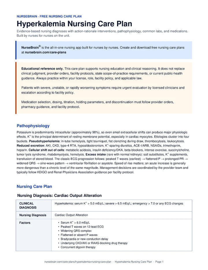
Nursing Care Plan
Nursing Diagnosis 1: Impaired Cardiac Output
Cardiac Output Alteration related to Hyperkalemia: serum K+ > 5.0 mEq/L; severe > 6.5 mEq/L; emergency > 7.0 or any ECG changes as evidenced by Serum K+ > 6.0 mEq/L; Peaked T waves on 12-lead ECG; Widening QRS complex; Flattened or absent P waves; Bradycardia or new conduction delay.
Interventions
- Place patient on continuous cardiac telemetry per facility protocol; obtain a 12-lead ECG on admission and after each K+-lowering intervention as ordered.
- Recheck serum K+ at intervals matched to clinical acuity and provider order during active treatment.
- Assess each ECG strip for peaked T waves, P-wave flattening, PR prolongation, and QRS widening; report findings promptly.
- Monitor HR, BP, and rhythm at intervals matched to clinical acuity and facility protocol during acute treatment.
- Review the most recent digoxin level and home medication list for ACE-I/ARB, K+-sparing diuretic, NSAIDs, TMP, and heparin; report findings to the provider team.
- On any K+ result > 6.0, verify draw technique with the team: tourniquet pressure, fist clenching, and whether the sample appeared hemolyzed; report findings.
- Administer IV calcium gluconate as ordered for K+ > 6.5 with ECG changes; in suspected digoxin toxicity, expect the provider to order a slower infusion (commonly over 20–30 min) and digoxin-Fab if available per facility protocol.
- Administer regular insulin and D50 as ordered per facility protocol; monitor glucose at the intervals ordered (commonly q15–30 min for the first 2 hours, then q1h through hour 6).
- Administer albuterol nebulizer as ordered per facility protocol.
- Confirm with the provider which home medications are held: ACE-I, ARB, K+-sparing diuretic, NSAIDs, TMP, and any K+ supplements.
- Administer SPS (Kayexalate), patiromer, or sodium zirconium cyclosilicate as ordered per facility protocol; flag concerns about ileus or recent surgery to the provider before giving SPS.
- Coordinate with nephrology and the provider team to prepare for emergency hemodialysis when K+ is refractory to medical therapy, with anuric AKI, or in ESRD on dialysis, per facility protocol.
- Educate the patient and family on the rationale for telemetry, repeat labs, and frequent finger-sticks in plain language.
- Teach the patient which home medications were held and that they should not be resumed without provider clearance.
- Educate the patient and family on a lower-K+ diet within the patient’s preferences and culture (commonly limiting bananas, oranges and OJ, potatoes, tomatoes, dried fruit, beans, and salt substitutes), per provider direction and dietitian recommendation.
- Notify the provider promptly for new widened QRS, sine-wave pattern, bradycardia below ordered parameters, or any pulseless rhythm; activate the code team per facility protocol.
- Coordinate nephrology consultation per facility protocol for refractory hyperkalemia or recurrent episodes.
Outcome: Serum K+ trend, ECG findings, and rhythm are monitored and reported per facility protocol; Resolution or improvement of peaked T waves and QRS width is documented on serial ECG; Sinus rhythm on continuous telemetry is monitored and changes are reported.
Nursing Diagnosis 2: Spiritual Distress
Electrolyte Imbalance related to Hyperkalemia: serum K+ > 5.0 mEq/L; severe > 6.5 mEq/L; emergency > 7.0 or any ECG changes as evidenced by Generalized muscle weakness; Patient reports of heavy or weak legs and difficulty rising from a chair; Reduced grip strength bilaterally; Paresthesias of the extremities; Sluggish or absent deep tendon reflexes.
Interventions
- Perform a baseline neuromuscular assessment: grip strength, lower-extremity strength on the 0–5 scale, DTRs, and paresthesias.
- Reassess motor strength and reflexes at intervals matched to clinical acuity and facility protocol during active treatment.
- Complete and document a fall-risk assessment on admission and per facility protocol.
- Ask the patient about subjective heaviness, fatigue, or difficulty with ADLs.
- Implement the fall-prevention bundle per facility protocol: bed alarm, non-skid socks, call light within reach, uncluttered room, and the appropriate assist level for transfers.
- Assist with ADLs and ambulation per facility protocol; use a gait belt and standby assist until strength returns toward baseline.
- Provide rest periods between activities; cluster cares to support uninterrupted recovery per facility protocol.
- Reposition every 2 hours when the patient is in bed and provide pressure-injury prevention per facility protocol.
- Teach the patient to call for help before getting out of bed or chair until cleared by the provider team.
- Educate the patient on the relationship between K+ level and muscle weakness, including that symptoms typically improve as labs normalize.
- Demonstrate safe transfer techniques and assistive device use before discharge, in coordination with PT when consulted.
- Notify the provider for new or worsening weakness despite improving K+, new paralysis, or respiratory-muscle involvement.
- Coordinate a physical therapy consult per facility protocol if weakness persists beyond several days of K+ correction.
Outcome: Muscle strength, reflexes, and paresthesias are monitored and changes are reported per facility protocol; Patient ambulates within ordered activity parameters with appropriate assistance; Fall-prevention bundle is implemented and documented per facility protocol.
Nursing Diagnosis 3: Non Adherence
Knowledge Deficit related to Hyperkalemia: serum K+ > 5.0 mEq/L; severe > 6.5 mEq/L; emergency > 7.0 or any ECG changes as evidenced by Patient verbalizes uncertainty about dietary K+ sources; Patient reports using salt substitutes (KCl-based); Patient is uncertain about which home medications to resume; First episode of hyperkalemia or a new CKD diagnosis; Limited prior education on lab-value targets.
Interventions
- Assess baseline knowledge of dietary K+, the kidney’s role, and current medications.
- Assess literacy, language, and learning preferences (visual, verbal, written) per facility protocol.
- Identify patient-stated barriers: cost, food access, polypharmacy, cognition, caregiver involvement.
- Coordinate with the dietitian for in-person lower-K+ diet teaching, with printed culturally relevant food lists per facility protocol.
- Provide a written medication list distinguishing "hold," "resume," and "changed dose" entries per provider order and facility discharge protocol.
- Use teach-back for each major topic: diet, medications, when to call, and follow-up labs.
- Teach high-K+ foods commonly limited in this context (such as bananas, oranges and OJ, potatoes, tomatoes, beans, dried fruit, avocado, spinach, and salt substitutes), within the patient’s preferences and culture and per dietitian recommendation.
- Specifically discuss KCl-based salt substitutes (such as Lite Salt, Nu-Salt, AlsoSalt) as a common, often-missed source of K+.
- Educate the patient and family on early warning signs (muscle weakness, palpitations, tingling, fatigue) and the importance of seeking care without delay.
- Teach the importance of follow-up K+ and Cr labs after discharge per the provider’s plan.
- Educate on hydration and on avoiding NSAIDs without provider clearance.
- Encourage the patient to discuss with the provider whether long-term ACE-I/ARB/MRA can be resumed alongside a K+ binder (such as patiromer or Lokelma) rather than stopped indefinitely.
- Coordinate follow-up with primary care or nephrology per provider order and facility discharge protocol.
- Refer to social work or community resources for food access if cost or availability is a stated barrier, per facility protocol.
Outcome: Patient identifies several high-K+ foods to limit per dietitian and provider recommendations; Patient verbalizes which medications were held and the plan for resumption; Patient states when to seek emergent care.
Nursing Diagnosis 4: Fluid Imbalance
Fluid Volume Alteration related to Hyperkalemia: serum K+ > 5.0 mEq/L; severe > 6.5 mEq/L; emergency > 7.0 or any ECG changes as evidenced by Underlying AKI or CKD; Concurrent IV therapy (calcium, insulin and dextrose, bicarbonate); Sodium-containing K+ binders (SPS, ZS-9); Diuretic therapy as adjunct treatment; Possible oliguria or anuria.
Interventions
- Maintain strict intake and output; calculate net balance at the intervals ordered (commonly every 8 hours) per facility protocol.
- Weigh the patient daily at the same time, on the same scale, in the same garments when clinically feasible.
- Auscultate lung fields at intervals matched to clinical acuity and facility protocol; assess for crackles, JVD, and peripheral edema.
- Trend BUN, Cr, and GFR with serial labs as ordered.
- Monitor for hypotension, tachycardia, and dry mucous membranes as signs that may reflect volume depletion from aggressive diuresis.
- Track all fluid administered for each K+-lowering intervention; document on the flowsheet per facility protocol.
- Use the minimum effective IV volumes per provider order; coordinate with pharmacy for appropriate concentrations.
- Administer loop diuretic (such as furosemide) as ordered per facility protocol when the provider team determines the kidneys are responsive.
- Coordinate early with nephrology and dialysis services per facility protocol when the patient is anuric or fluid overload is not responding to medical therapy.
- Teach the patient daily weights at home and which thresholds the provider has identified for calling in (commonly more than 2 lb overnight or 5 lb in a week, per provider order).
- Educate on the prescribed fluid plan when fluid restriction is ordered, including practical tools (such as a marked container) per facility protocol.
- Review signs of fluid overload (worsening SOB, leg swelling, weight gain) and dehydration (dizziness, dark urine, dry mouth).
- Notify the provider for UOP below ordered parameters, new pulmonary congestion, or rapid weight gain.
- Coordinate nephrology and dialysis-access planning per facility protocol when recurrent hyperkalemia and progressive CKD are part of the clinical picture.
Outcome: Net intake and output are monitored and reported within ordered parameters per facility protocol; Urine output is monitored and reported (target generally ≥ 0.5 mL/kg/hr when non-anuric, per order); Pulmonary findings are monitored and worsening is reported promptly.
Nursing Diagnosis 5: Anxiety
Anxiety related to Hyperkalemia: serum K+ > 5.0 mEq/L; severe > 6.5 mEq/L; emergency > 7.0 or any ECG changes as evidenced by Patient verbalization of fear about cardiac arrest or the unknown; Restlessness or hypervigilance disproportionate to the clinical state; Rapid pace of treatment witnessed by patient and family (multiple lines, frequent labs); Unfamiliarity with hyperkalemia, telemetry, and ICU- or stepdown-level monitoring; Sleep disturbance from monitor alarms and frequent interventions.
Interventions
- Assess anxiety level using a 0–10 scale at the start of every shift and PRN per facility protocol.
- Identify the patient’s stated triggers (such as monitor alarms, repeat finger-sticks, fear of cardiac arrest, fear of family separation).
- Observe for physical signs of anxiety: tachycardia out of proportion to clinical state, restlessness, hand-wringing, hypervigilance.
- Assess sleep quality and contributors daily (such as alarms, lighting, frequent interventions, anxiety).
- Provide a calm, reassuring presence; speak clearly and at a measured pace.
- Explain procedures and findings in plain, patient-friendly terms before performing them.
- Cluster cares to support uninterrupted rest periods when clinical state allows per facility protocol.
- Limit non-essential nighttime stimuli (such as overhead lights, loud conversations, unnecessary alarms) per facility protocol.
- Facilitate family presence and contact within unit policy, including video or phone when in-person is restricted.
- Coordinate with chaplaincy, social work, or interpreter services per patient preference and facility policy.
- Teach diaphragmatic breathing and grounding techniques the patient can use independently.
- Educate the patient and family on hyperkalemia in plain language: what K+ does, why the treatment cascade looks busy, and what improvement looks like on telemetry and labs.
- Teach the family what each monitor alarm means and what is and is not clinically concerning.
- Coordinate with chaplaincy, social work, or psych services per facility protocol if anxiety persists or worsens despite non-pharmacologic measures.
- Notify the provider for severe or persistent anxiety unresponsive to non-pharmacologic measures.
Outcome: Patient verbalizes decreased anxiety; Patient demonstrates at least one coping strategy (such as diaphragmatic breathing or grounding); Patient sleeps in longer blocks when clinical state allows.
Pathophysiology
Potassium is predominantly intracellular (approximately 98%), so even small extracellular shifts can produce major physiologic effects. K+ is the principal determinant of resting membrane potential, especially in cardiac myocytes. Etiologies cluster into four buckets. Pseudohyperkalemia: in-tube hemolysis, tight tourniquet, fist clenching during draw, thrombocytosis, leukocytosis. Reduced excretion: AKI, CKD, type-4 RTA, hypoaldosteronism, K+-sparing diuretics, ACE-I/ARB, NSAIDs, trimethoprim, heparin. Cellular shift out of cells: metabolic acidosis, insulin deficiency/DKA, beta-blockers, intense exercise, succinylcholine, tumor lysis syndrome, rhabdomyolysis, hemolysis. Excess intake (rare with normal kidneys): salt substitutes, K+ supplements, transfusion of stored blood. The classic ECG progression follows: peaked T waves (earliest) → flattened P → prolonged PR → widened QRS → sine-wave pattern → ventricular fibrillation or asystole. Speed of rise matters; an acute increase is generally more dangerous than a chronic level of the same magnitude. Management decisions are coordinated by the provider team and typically follow KDIGO and Renal Physicians Association guidance per facility protocol.
Quick Reference
- Treatment threshold: K+ ≥ 6.0 mEq/L or any ECG change
- SPS (Kayexalate) caution: Colonic necrosis risk (FDA boxed warning)
- Newer K+ binders: Patiromer, ZS-9 available where ordered
- Insulin + dextrose dose: 10 U regular insulin + 25 g D50 IV per order
- Calcium gluconate: Membrane stabilization; does not lower K+
Common Labs
| Lab | Normal range | Significance in Hyperkalemia |
|---|---|---|
| Serum K+ | 3.5–5.0 mEq/L | Nurses support a free-flowing draw, avoid fist clenching, and deliver promptly to reduce in-tube hemolysis. Repeat draw is commonly requested by the provider when the result is unexpected. |
| ECG (12-lead + tele) | Normal complexes | Peaked Ts, flat P, wide QRS, and sine wave are reported to the provider promptly. Even subtle peaked Ts can prompt the provider team to begin treatment per facility protocol. |
| BUN / Cr | 7–20 / 0.6–1.2 mg/dL | Reduced excretion is a leading cause; nurses trend renal function and report worsening values. |
| HCO3− / pH (ABG) | 22–26 mEq/L / 7.35–7.45 | Acidosis can drive K+ out of cells. Trend supports provider-team decisions about correcting the underlying acidosis. |
| Glucose | 70–110 mg/dL | Baseline before insulin and dextrose is ordered; hypoglycemia is most common 1–6 hours post-dose and is monitored closely. |
| Mg2+ | 1.7–2.2 mg/dL | Often coexists with K+ disturbance; the provider may order repletion per facility protocol. |
| Digoxin level | 0.8–2.0 ng/mL | Digoxin can amplify K+-related cardiac toxicity. Nurses report the level promptly. Per current evidence (Lindner 2020), IV calcium is not withheld for life-threatening hyperkalemic ECG changes; the provider team may order a slower infusion (over 20–30 min) and digoxin-Fab if available per facility protocol. |
| CPK | < 200 U/L | Rhabdomyolysis can release intracellular K+; nurses trend and report rising values. |
| LDH / haptoglobin | LDH < 250 / Hapto 30–200 | Help support assessment of hemolysis as a contributor; reported to the provider team. |
| Uric acid / phosphate | UA < 7 / PO4 2.5–4.5 | Support tumor-lysis-syndrome screening when chemotherapy or hematologic malignancy is in the clinical picture per provider order. |
Common Medications
| Class | Examples | Mechanism of action | Key side effects | Nursing considerations |
|---|---|---|---|---|
| IV calcium gluconate | Calcium gluconate 1–2 g IV per order | Raises threshold potential and restores myocyte excitability; membrane stabilization, does not lower K+. | Tissue necrosis on extravasation, hypercalcemia; the historic "stone heart" concern with digoxin toxicity is based on weak evidence and IV calcium is not withheld for life-threatening hyperkalemic ECG changes. | Administer as ordered per provider direction, pharmacy guidance, and facility protocol. In suspected digoxin toxicity, the provider may order a slower infusion (commonly over 20–30 min) and digoxin-Fab if available. Onset is typically within minutes; effect commonly lasts 30–60 minutes. Nurses monitor ECG, IV site for extravasation, and report persistent ECG changes for possible redose per order. |
| Insulin + dextrose | Regular insulin 10 U IV + 25 g D50 IV per order | Insulin drives K+ into cells via Na+/K+-ATPase. | Hypoglycemia (commonly 15 min and 1–6 h post-dose); over-correction to hypokalemia. | Administer as ordered per provider direction and facility protocol. Onset is typically within 15 minutes; effect can last 4–6 hours. Nurses monitor glucose q15–30 min for the first 2 hours, then q1h through hour 6 per facility protocol. In CKD/ESRD the provider may order a D10W infusion (commonly 50–75 mL/h) after the D50 bolus to reduce delayed hypoglycemia. |
| Beta-2 agonist | Albuterol nebulizer 10–20 mg per order | β2 stimulation activates Na+/K+-ATPase and supports intracellular shift. | Tachycardia, tremor, hyperglycemia; approximately 25% of patients respond poorly. | Administer as ordered per provider direction and facility protocol. Often synergistic with insulin; effect onset is commonly about 30 minutes. Generally not selected as monotherapy by the provider team. |
| Sodium bicarbonate | NaHCO3 IV per order | Correcting acidosis can support intracellular K+ movement. | Hypernatremia, volume overload, alkalosis. | Administer as ordered per provider direction and facility protocol. Commonly more useful when frank acidosis is present; selection and dose are provider-team decisions. |
| Sodium polystyrene sulfonate | SPS (Kayexalate) PO/PR per order | Cation-exchange resin that binds K+ in the colon. | FDA boxed warning: colonic necrosis; constipation, hypernatremia, hypocalcemia. | Administer as ordered per provider direction, pharmacy guidance, and facility protocol. Caution is generally advised in ileus, recent surgery, or opioid-related slowed gut transit; the provider team may select a newer binder instead per facility protocol. |
| Patiromer | Veltassa PO daily per order | Binds K+ in the colon via calcium exchange. | Constipation, hypomagnesemia, edema. | Administer as ordered per provider direction and facility protocol. Onset is slower (commonly several hours); pharmacy guidance typically supports separating from other oral medications by about 3 hours. |
| Sodium zirconium cyclosilicate | Lokelma (ZS-9) PO per order | Selective K+ binder via cation exchange in the GI tract. | Edema, hypokalemia if over-corrected, QT prolongation. | Administer as ordered per provider direction and facility protocol. Faster onset (commonly about 1 hour); may be selected acutely by the provider team. |
| Hemodialysis | HD or CRRT per provider order | Removes K+ directly from the blood. | Hypotension, access complications, dialysis disequilibrium. | Administer as ordered. Definitive therapy decisions (when medical therapy is not lowering K+, with anuria, or in ESRD on dialysis) are coordinated by the provider team per facility protocol. Rebound K+ can occur post-HD; nurses trend the value and report rising trends. |
References
- Makic, M. B. F., & Martinez-Kratz, M. R. (Eds.). (2023). Ackley and Ladwig’s Nursing Diagnosis Handbook: An Evidence-Based Guide to Planning Care (13th ed.). Elsevier.
- Palmer, B. F., & Clegg, D. J. (2017). Diagnosis and Treatment of Hyperkalemia. Mayo Clinic Proceedings, 92(10), 1546–1554.
- Lindner, G., Burdmann, E. A., Clase, C. M., et al. (2020). Acute Hyperkalemia in the Emergency Department: A Summary from a Kidney Disease: Improving Global Outcomes Conference. Journal of Emergency Medicine, 59(1), 43–54.
Frequently Asked Questions
What is the nursing care plan for Hyperkalemia?
A Hyperkalemia nursing care plan organizes the assessment, nursing diagnoses, goals, interventions, and evaluation criteria for a patient with Hyperkalemia. Diagnoses are ordered by what is currently most destabilizing for the patient.
What are the priority nursing diagnoses for Hyperkalemia?
Priority diagnoses for Hyperkalemia appear in the Nursing Diagnoses section above, ordered by clinical acuity. The top diagnosis should reflect what is currently most destabilizing for this specific patient.
What is the priority nursing intervention for Hyperkalemia?
Priority interventions for Hyperkalemia are listed in the care plan above, organized by diagnosis. The most critical actions address airway, circulation, and the highest-acuity problem first.
What complications should the nurse monitor for in Hyperkalemia?
Complications to monitor for in Hyperkalemia are listed within each diagnosis section above. Trend vitals, mental status, and the condition-specific red flags described in the assessment section.