Asthma Exacerbation Nursing Care Plan
Asthma exacerbation nursing care plan with prioritized diagnoses, bronchodilator administration, breathing-pattern monitoring, and a printable PDF.
Nursing Care Plan
Nursing Diagnosis 1: Impaired Breathing
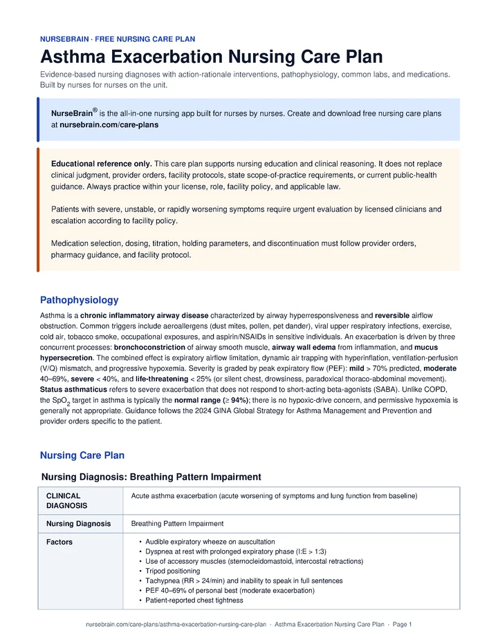
Breathing Pattern Impairment related to Acute asthma exacerbation (acute worsening of symptoms and lung function from baseline) as evidenced by Audible expiratory wheeze on auscultation; Dyspnea at rest with prolonged expiratory phase (I:E > 1:3); Use of accessory muscles (sternocleidomastoid, intercostal retractions); Tripod positioning; Tachypnea (RR > 24/min) and inability to speak in full sentences.
Interventions
- Auscultate all lung fields at intervals matched to clinical acuity and facility protocol during acute exacerbation; document presence, location, and timing (inspiratory vs expiratory) of wheeze.
- Measure PEF before and after each ordered bronchodilator treatment per facility protocol; compare to personal best.
- Count respiratory rate over a full 60 seconds; observe depth, symmetry, and accessory-muscle use at intervals matched to clinical acuity.
- Assess ability to speak in complete sentences; document sentence length (full / phrases / single words / unable).
- Monitor for findings consistent with impending respiratory failure: drowsiness, confusion, silent chest, paradoxical breathing, PaCO2 rising from low or normal toward mid-40s.
- Support the patient in high-Fowler’s or tripod position per tolerance; avoid forcing supine.
- Administer ordered SABA (albuterol) via nebulizer or MDI plus spacer per provider order and facility protocol (commonly q20 minutes × 3 doses, then PRN).
- Administer ordered ipratropium (SAMA) as an add-on to SABA nebulizer in moderate-to-severe exacerbations per provider direction and facility protocol.
- Administer ordered systemic corticosteroid (prednisone 40–50 mg PO or methylprednisolone IV) early in the encounter per provider direction and facility protocol.
- For severe exacerbation that does not respond to initial therapy, prepare and administer ordered magnesium sulfate 2 g IV over 20 minutes per provider direction and facility protocol.
- Anticipate and prepare for possible intubation and non-invasive ventilation per facility protocol when clinical trajectory raises concern: confirm airway cart at bedside, draw induction medications per provider order, and request the senior airway operator early.
- Demonstrate correct MDI plus spacer technique: shake, exhale, seal lips on spacer, slow deep inhalation, hold breath about 10 seconds, wait 30–60 seconds between puffs.
- Teach pursed-lip and diaphragmatic breathing techniques for acute dyspnea management.
- Educate on the distinction between reliever (SABA, for symptoms) and controller (ICS or ICS-LABA, daily even when well).
- Provide and review a written Asthma Action Plan (AAP) per facility protocol: green / yellow / red zones with PEF cutoffs and specific actions.
- Teach the patient to rinse the mouth after every ICS dose to support prevention of oral candidiasis and dysphonia.
- Notify the provider for PEF < 50% of personal best despite treatment, SpO2 below ordered parameters on supplemental O2, rising PaCO2, or any sign of impending failure.
- Coordinate respiratory therapy and ICU evaluation early for severe or refractory exacerbations per facility protocol.
Outcome: Respiratory rate trends toward 12–20 breaths/min at rest within ordered parameters; Wheeze is monitored and reported; resolution or marked diminution is documented when present; PEF trend is monitored and reported; improvement toward ≥ 80% of personal best is supported.
Nursing Diagnosis 2: Impaired Gas Exchange
Gas Exchange Impairment related to Acute asthma exacerbation (acute worsening of symptoms and lung function from baseline) as evidenced by SpO2 < 94% on room air; ABG: pH 7.46 / PaO2 62 mmHg / PaCO2 32 mmHg (early; respiratory alkalosis); V/Q mismatch from airway obstruction and air trapping; Restlessness, anxiety, and tachycardia; Cyanosis of lips or nail beds (late sign).
Interventions
- Apply continuous pulse oximetry; document SpO2 trend at intervals matched to clinical acuity and facility protocol.
- Obtain ordered ABG for severe exacerbation, SpO2 below ordered parameters on O2, or any clinical deterioration per provider order and facility protocol.
- Monitor for early findings that may reflect hypoxia (restlessness, anxiety, tachycardia) and late findings (cyanosis, bradycardia, decreased LOC).
- Assess capillary refill, skin color, and nail-bed perfusion at intervals matched to clinical acuity.
- Monitor heart rate, rhythm, and BP continuously during high-dose SABA therapy.
- Administer ordered supplemental O2 via nasal cannula or simple mask, titrated within provider parameters to support SpO2 94–98%.
- Support high-Fowler’s positioning; if tolerated, encourage forward lean with arms supported on an overbed table.
- Administer ordered hydration (oral or IV) to support secretion clearance; avoid fluid overload.
- Coach the patient through each ordered nebulizer treatment: slow deep breath in, hold, slow exhale; provide reassurance during treatment.
- Prepare for non-invasive ventilation (BiPAP) or intubation if hypercapnic respiratory failure develops, per provider direction and facility protocol.
- Educate on the difference between the typical asthma SpO2 target (≥ 94%) and any prior COPD or CHF instructions the patient may have received.
- Teach findings of worsening gas exchange that should prompt calling 911: lips or nails turning blue, confusion, drowsiness, inability to speak.
- Educate on smoking cessation and avoidance of secondhand smoke; offer cessation resources per facility protocol.
- Notify the provider for SpO2 below ordered parameters on supplemental O2, PaCO2 rising into normal range in a tachypneic patient, or any decreased LOC.
- Coordinate ICU transfer for findings consistent with impending respiratory failure: silent chest, drowsiness, paradoxical breathing, or PEF < 25%, per facility protocol.
Outcome: Patient maintains SpO2 within ordered parameters on room air or the lowest supplemental O2 that meets the goal; ABG findings, including PaO2 and PaCO2 trend, are monitored and reported per facility protocol; Patient reports decreased air hunger when clinical state permits.
Nursing Diagnosis 3: Impaired Urinary System Function
Anxiety related to Acute asthma exacerbation (acute worsening of symptoms and lung function from baseline) as evidenced by Patient verbalization of fear of suffocation or dying; Visible apprehension, hand-wringing, wide eyes; Tachycardia and tachypnea disproportionate to clinical state; Hyperventilation that can worsen dynamic air trapping; Previous near-fatal exacerbation or ICU admission.
Interventions
- Assess anxiety on a 0–10 scale at the start of every shift and during each dyspnea episode.
- Identify the patient’s specific triggers for anxiety (sensation of suffocation, prior ICU stay, monitor alarms).
- Observe for physical signs: tachycardia out of proportion to PEF, restlessness, hyperventilation, panic facies.
- Provide a calm, confident presence during acute dyspnea episodes.
- Speak slowly, in short clear sentences; model the slow breathing pattern you want the patient to adopt.
- Coach pursed-lip breathing in real time during each acute episode; count inhalations and exhalations aloud.
- Cluster care and protect rest periods of at least 30 minutes between treatments when clinical state allows.
- Explain every procedure, alarm, and finding in plain language before acting.
- Teach the cycle: panic → hyperventilation → air trapping → worse dyspnea → more panic; and how slow breathing can help break the cycle.
- Practice diaphragmatic breathing, pursed-lip breathing, and a grounding exercise (e.g., 5-4-3-2-1 senses).
- Discuss the Asthma Action Plan as a concrete sense of control: green/yellow/red zones with pre-decided actions.
- Educate family on supportive presence: calm voice, no crowding, validating fear without amplifying it.
- Coordinate social work or behavioral-health referral per facility protocol for patients with prior near-fatal exacerbation or persistent anxiety.
- Notify the provider for severe anxiety unresponsive to non-pharmacologic measures; anxiolytics are used cautiously in this population due to respiratory-depressant risk.
Outcome: Patient verbalizes decreased anxiety using a 0–10 scale; Patient demonstrates pursed-lip or diaphragmatic breathing during dyspnea episodes; Heart rate and respiratory rate remain consistent with clinical state, not panic-driven.
Nursing Diagnosis 4: Non Adherence
Knowledge Deficit related to Acute asthma exacerbation (acute worsening of symptoms and lung function from baseline) as evidenced by New asthma diagnosis or first severe exacerbation; Verbalized confusion about reliever vs controller inhalers; Inability to demonstrate correct MDI technique; No written Asthma Action Plan; Recurrent ED visits or hospitalizations.
Interventions
- Assess current understanding of asthma, medications, and triggers using open-ended questions.
- Have the patient demonstrate inhaler technique before teaching; document specific errors.
- Review prior controller adherence: prescription refill history, days-on-therapy, patient self-report.
- Identify the patient’s individual triggers through history: allergens, viral URI, exercise, cold air, smoke, NSAIDs, occupational exposures.
- Assess health literacy, preferred learning style, and language preferences.
- Provide written materials at an appropriate reading level in the patient’s preferred language per facility protocol.
- Use the teach-back method for every key topic: ask the patient to explain it in their own words.
- Provide and review a personalized written Asthma Action Plan with PEF or symptom-based zones per facility protocol.
- Coordinate spacer prescription and demonstration before discharge if the patient uses MDIs.
- Explain the difference between reliever (SABA, fast onset, for symptoms) and controller (ICS or ICS-LABA, slow, daily, prevents flares).
- For patients managed under GINA Track 1, explain that as-needed low-dose ICS-formoterol can be the preferred Step 1–2 reliever (replacing SABA-only); under NAEPP / US-based plans, SABA-PRN can remain acceptable at Step 1.
- Demonstrate correct MDI plus spacer technique step by step; have the patient return-demonstrate; repeat until correct.
- Teach trigger identification and avoidance: dust-mite covers, HEPA filters, pet-free bedroom, no smoking indoors, pre-exercise SABA for EIB.
- Review the AAP zones: green (PEF > 80%, no symptoms, continue controller); yellow (PEF 50–79% or mild symptoms, add SABA ± oral steroid per plan); red (PEF < 50% or severe symptoms, SABA plus steroid plus 911).
- Educate on annual influenza vaccination and pneumococcal vaccination per CDC recommendations and provider order.
- Teach when to seek emergency care: no SABA response, inability to speak in sentences, blue lips or nails, drowsiness, or PEF < 50%.
- Coordinate primary care or pulmonology follow-up within 1–4 weeks of discharge per facility protocol.
- Refer to an asthma educator, pulmonary rehab, or community asthma program when available per facility protocol.
Outcome: Patient correctly identifies controller and reliever medications by name and purpose; Patient demonstrates correct MDI plus spacer technique on teach-back; Patient verbalizes personal triggers and at least one avoidance strategy for each.
Nursing Diagnosis 5: Activity Intolerance
Activity Intolerance related to Acute asthma exacerbation (acute worsening of symptoms and lung function from baseline) as evidenced by History of exercise-induced bronchospasm (EIB); Dyspnea on exertion at less than usual activity level; PEF drops > 15% from pre-activity baseline after exertion; Patient avoidance of physical activity from fear of symptoms; Deconditioning from sedentary lifestyle.
Interventions
- Monitor vital signs, SpO2, and PEF before, during, and after activity per facility protocol.
- Assess patient-reported dyspnea on a 0–10 scale before and after activity.
- Identify activities or exposures (cold air, exercise, allergen-rich environments) that consistently trigger symptoms.
- Observe for findings consistent with exercise-induced bronchospasm: wheezing, cough, chest tightness 5–15 minutes after starting exercise.
- Administer ordered SABA prior to known triggers (exercise, cold-air exposure) per provider direction and facility protocol.
- Coordinate ambulation and PT sessions during peak bronchodilator effect (approximately 30–60 minutes after a SABA dose).
- Provide rest periods between activities; cluster care to allow uninterrupted rest blocks.
- Coordinate physical therapy for a graded reconditioning program per provider order.
- Teach that good asthma control generally allows normal activity, including exercise.
- Educate on pre-exercise SABA use, warm-up periods, and avoidance of cold-air exercise when possible.
- Teach energy-conservation: pace activities, prioritize tasks, sit while performing tasks when possible.
- Educate on regular aerobic activity (walking, swimming) as part of long-term asthma control.
- Teach the patient to monitor PEF before and after activity to track personal tolerance and identify deterioration.
- Coordinate referral to pulmonary rehabilitation when deconditioning is severe per facility protocol.
- Notify the provider for persistent EIB despite pre-treatment or for new exertional chest pain.
Outcome: Patient ambulates within ordered parameters without significant dyspnea or PEF drop; Patient pre-medicates with ordered SABA prior to known triggers when indicated; PEF drop with activity is monitored and reported; trend toward < 15% of personal best is supported.
Pathophysiology
Asthma is a chronic inflammatory airway disease characterized by airway hyperresponsiveness and reversible airflow obstruction. Common triggers include aeroallergens (dust mites, pollen, pet dander), viral upper respiratory infections, exercise, cold air, tobacco smoke, occupational exposures, and aspirin/NSAIDs in sensitive individuals. An exacerbation is driven by three concurrent processes: bronchoconstriction of airway smooth muscle, airway wall edema from inflammation, and mucus hypersecretion. The combined effect is expiratory airflow limitation, dynamic air trapping with hyperinflation, ventilation-perfusion (V/Q) mismatch, and progressive hypoxemia. Severity is graded by peak expiratory flow (PEF): mild > 70% predicted, moderate 40–69%, severe < 40%, and life-threatening < 25% (or silent chest, drowsiness, paradoxical thoraco-abdominal movement). Status asthmaticus refers to severe exacerbation that does not respond to short-acting beta-agonists (SABA). Unlike COPD, the SpO2 target in asthma is typically the normal range (≥ 94%); there is no hypoxic-drive concern, and permissive hypoxemia is generally not appropriate. Guidance follows the 2024 GINA Global Strategy for Asthma Management and Prevention and provider orders specific to the patient.
Quick Reference
- SpO2 target: ≥ 94% (not 88–92%)
- PEF severity: >70% mild · 40–69% mod · <40% severe
- Albuterol neb: 2.5–5 mg q20min × 3 doses
- Systemic steroid: Prednisone 40–50 mg × 5 days
- Magnesium sulfate: 2 g IV over 20 min (severe)
Common Labs
| Lab | Normal range | Significance in Asthma |
|---|---|---|
| Peak Expiratory Flow (PEF) | > 80% personal best (or predicted if no baseline) | Bedside trend used to support provider-team decisions; for newly diagnosed patients, height/age/sex-predicted values are commonly used until 2 weeks of green-zone tracking establishes personal best. < 40% predicted is consistent with severe exacerbation; < 25% can indicate life-threatening exacerbation and should prompt urgent reassessment and provider notification. |
| ABG | pH 7.35–7.45 / PaCO2 35–45 | A rising PaCO2 in a tachypneic asthmatic can signal fatigue and impending respiratory failure rather than stabilization; nurses report the trend to the provider team promptly. |
| SpO2 | ≥ 94% on RA | Goal is generally the normal range; oxygen is titrated to keep SpO2 ≥ 94% (93–95% in pregnancy) per provider order and facility protocol. |
| CBC with diff | Eos < 500 cells/µL | Eosinophilia can support a type-2/allergic phenotype assessment by the provider team and may inform biologic-therapy decisions during outpatient follow-up. |
| Total IgE | < 100 IU/mL | Elevated in atopic asthma; reported to the provider team to support outpatient decisions about omalizumab dosing per specialist evaluation. |
| CXR | Clear lung fields | Not routinely obtained; ordered by the provider team to help rule out pneumothorax, pneumonia, or foreign body when clinical findings raise concern. |
| Theophylline level | 5–15 µg/mL | Rarely used in current practice; narrow therapeutic window. Monitored per provider order when the patient is on chronic theophylline. |
| CMP (K+) | 3.5–5.0 mEq/L | High-dose SABA and systemic steroids can cause hypokalemia; nurses trend potassium and administer ordered replacement per facility electrolyte-replacement protocol to support arrhythmia prevention. |
| Respiratory pathogen panel | Negative | Viral URI (rhinovirus, RSV, influenza) is the most common trigger; results can inform isolation precautions per facility infection-prevention policy. |
| FeNO (fractional exhaled NO) | < 25 ppb | Outpatient marker of eosinophilic airway inflammation; results support provider-team decisions about ICS step-up during follow-up. |
Common Medications
| Class | Examples | Mechanism of action | Key side effects | Nursing considerations |
|---|---|---|---|---|
| SABA (short-acting β2-agonist) | Albuterol, Levalbuterol | Stimulates β2 receptors → bronchial smooth-muscle relaxation; commonly first-line rescue per GINA 2024 and NAEPP EPR-4 (2020). | Tachycardia, tremor, hypokalemia, hyperglycemia, lactic acidosis (high-dose). | Administer as ordered. Nebulized 2.5–5 mg q20min × 3, then PRN, or continuous 10–15 mg/hr in severe exacerbation per provider order and facility protocol. MDI plus spacer can be equivalent to a nebulizer in a cooperative patient when ordered. Nurses monitor HR, rhythm, tremor, potassium trend, and bronchodilator response (PEF, work of breathing, breath sounds); escalate new tachyarrhythmia or absent response per facility protocol. |
| SAMA (anticholinergic) | Ipratropium (Atrovent), DuoNeb (with albuterol) | Blocks muscarinic M3 receptors → bronchodilation; can be synergistic with SABA. | Dry mouth, urinary retention, blurred vision if eye exposure. | Administer as ordered as an add-on to SABA in moderate-to-severe ED exacerbations per provider direction and facility protocol; not typically used as monotherapy and offers little additional benefit after admission. Nurses monitor bronchodilator response and adverse effects. |
| Systemic corticosteroid | Prednisone PO, Methylprednisolone IV | Reduces airway inflammation and edema; supports restoration of β-receptor responsiveness; onset 4–6 h, so early administration matters. | Hyperglycemia, mood changes, insomnia, fluid retention, GI upset; a short course is generally associated with minimal adrenal suppression. | Administer as ordered per provider direction and facility protocol. Typical regimens include prednisone 40–50 mg PO daily × 5 days (no taper needed in most cases) or IV methylprednisolone 60–125 mg if the patient is NPO or severely ill. Nurses monitor glucose, mood, and GI tolerance, and reinforce the importance of completing the ordered course. |
| ICS (inhaled corticosteroid) | Fluticasone, Budesonide, Beclomethasone | Anti-inflammatory; reduces airway hyperresponsiveness; controller, not a rescue medication. | Oral candidiasis (rinse mouth after each dose), dysphonia, minimal systemic absorption. | Administer as ordered per provider direction. Commonly introduced or escalated at discharge as part of step-up therapy and Asthma Action Plan green-zone use; full effect can take weeks. Nurses reinforce ordered technique and rinse-and-spit after each dose. |
| ICS-LABA combination | Budesonide/Formoterol (Symbicort), Beclomethasone/Formoterol (SMART-eligible); Fluticasone/Salmeterol (Advair) (controller only) | Long-acting bronchodilation plus anti-inflammatory effect; SMART/MART therapy uses ICS-formoterol (rapid-onset LABA) as a single inhaler for maintenance plus reliever per GINA 2024; salmeterol-based combos are not used for SMART because of slow onset. | LABA carries a BLACK-BOX warning against use as monotherapy in asthma due to increased asthma-related death. | Administer as ordered as Step 3+ controller per GINA 2024 and facility protocol. Nurses reinforce that LABA is paired with ICS per the prescribed regimen and escalate any patient report of using LABA alone to the provider team. |
| Magnesium sulfate IV | Magnesium sulfate 2 g IV | Smooth-muscle relaxation via calcium-channel inhibition; anti-inflammatory; severe-exacerbation adjunct. | Hypotension, flushing, warmth, hyporeflexia at high levels; caution in renal impairment. | Administer as ordered, commonly 2 g IV over 20 minutes for severe exacerbation that does not respond to SABA plus systemic steroid, per provider direction and facility protocol. Nurses monitor BP, DTRs, respiratory effort, and rhythm; escalate hypotension or new hyporeflexia per facility protocol. |
| Biologics (severe persistent) | Omalizumab (anti-IgE), Mepolizumab/Benralizumab (anti-IL-5), Dupilumab (anti-IL-4Rα) | Targeted blockade of type-2 inflammatory pathways; can reduce exacerbations 50%+ in selected phenotypes. | Injection-site reaction, anaphylaxis (omalizumab; observe per facility protocol after dose), eosinophilia. | Outpatient long-term therapy, not used for acute exacerbation. Initiated by specialists after phenotype and biomarker selection. Nurses coordinate referral and reinforce ordered post-dose observation per facility protocol. |
| Heliox (helium-oxygen mix) | 70:30 or 80:20 He:O2 | Lower-density gas can reduce turbulent airflow resistance → decreased work of breathing. | Cannot be delivered if FiO2 requirement > 30% (limits helium fraction); voice change. | Administer as ordered as a last-resort bridge in severe refractory asthma to allow time for steroids to take effect; commonly ICU- and respiratory-therapy-driven per provider direction and facility protocol. Nurses monitor work of breathing, oxygenation, and patient tolerance; escalate worsening trajectory per protocol. |
| Epinephrine (impending respiratory failure) | Epinephrine 0.3–0.5 mg IM (1 mg/mL) | Non-selective adrenergic agonist; can support bronchodilation, reduce airway edema, and support hemodynamics when the patient is deteriorating toward arrest. | Tachyarrhythmia, hypertension, anxiety, tremor; use with caution in older adults and patients with cardiovascular disease. | Administer as ordered for impending respiratory failure or anaphylactoid component per provider direction and facility protocol (commonly IM to the lateral thigh). Nurses monitor rhythm, BP, and respiratory trajectory and escalate per protocol. |
| Ketamine (induction / sedation) | Ketamine IV per provider order | NMDA-receptor antagonist with bronchodilator and sympathomimetic properties; can be considered by the provider team for induction or sedation in severe refractory asthma. | Hypertension, tachycardia, emergence reactions, increased oral secretions; provider team weighs risks and benefits. | Administer as ordered per provider direction and facility protocol when ketamine is selected for induction during high-risk intubation or for sedation in severe refractory asthma. Nurses monitor hemodynamics, secretions, and emergence and document response. |
References
- Makic, M. B. F., & Martinez-Kratz, M. R. (Eds.). (2023). Ackley and Ladwig’s Nursing Diagnosis Handbook: An Evidence-Based Guide to Planning Care (13th ed.). Elsevier.
- Global Initiative for Asthma. (2024). Global Strategy for Asthma Management and Prevention. https://ginasthma.org
- National Asthma Education and Prevention Program Coordinating Committee Expert Panel Working Group. (2020). 2020 Focused Updates to the Asthma Management Guidelines (EPR-4). Journal of Allergy and Clinical Immunology, 146(6), 1217–1270.
- National Asthma Education and Prevention Program. (2007). Expert Panel Report 3 (EPR-3): Guidelines for the Diagnosis and Management of Asthma. National Heart, Lung, and Blood Institute.
Frequently Asked Questions
What is the nursing care plan for Asthma?
A Asthma nursing care plan organizes the assessment, nursing diagnoses, goals, interventions, and evaluation criteria for a patient with Asthma Exacerbation. Diagnoses are ordered by what is currently most destabilizing for the patient.
What are the priority nursing diagnoses for Asthma?
Priority diagnoses for Asthma appear in the Nursing Diagnoses section above, ordered by clinical acuity. The top diagnosis should reflect what is currently most destabilizing for this specific patient.
What is the priority nursing intervention for Asthma?
Priority interventions for Asthma are listed in the care plan above, organized by diagnosis. The most critical actions address airway, circulation, and the highest-acuity problem first.
What complications should the nurse monitor for in Asthma?
Complications to monitor for in Asthma are listed within each diagnosis section above. Trend vitals, mental status, and the condition-specific red flags described in the assessment section.
A report sheet built for this patient
This is exactly the kind of patient these NurseBrain report sheets are built for. Keep everything you track on one page, all shift.