COPD Exacerbation Nursing Care Plan
COPD exacerbation nursing care plan with prioritized diagnoses, airway clearance, gas exchange, and a printable PDF. Built by nurses for nurses.
Nursing Care Plan
Nursing Diagnosis 1: Impaired Airway Clearance
Airway Clearance Impairment related to Acute exacerbation of chronic obstructive pulmonary disease (AECOPD) as evidenced by Diffuse rhonchi and expiratory wheeze on auscultation; Increased sputum volume with purulent (yellow-green) appearance; Weak, ineffective cough with poor secretion clearance; Patient-reported chest tightness and difficulty expectorating; Prolonged expiratory phase, accessory-muscle use.
Interventions
- Auscultate breath sounds in all lung fields at intervals matched to clinical acuity and facility protocol; document location and quality of adventitious sounds.
- Assess respiratory rate, depth, effort, and accessory-muscle use at intervals matched to clinical acuity.
- Document sputum volume, color, consistency, and odor per shift and per facility protocol.
- Evaluate effectiveness of cough; note whether secretions are mobilized.
- Monitor SpO2 continuously per facility protocol; track the trend rather than single values.
- Administer scheduled and PRN bronchodilators (SABA + SAMA via DuoNeb) as ordered; reassess in 15–30 minutes per facility protocol.
- Position patient in high-Fowler’s or the tripod position (sitting forward, arms braced on overbed table) when tolerated.
- Encourage oral fluid intake (commonly 1.5–2 L/day) per provider order, unless contraindicated by cardiac or renal status.
- Coach the huff cough technique and provide tissues plus a labeled sputum cup at the bedside.
- Coordinate with respiratory therapy for chest physiotherapy or an oscillatory PEP device (for example, Acapella) per provider order and facility protocol.
- Teach pursed-lip breathing: inhale through the nose 2 counts, exhale through pursed lips 4 counts.
- Teach proper inhaler and spacer technique; have the patient demonstrate before discharge.
- Educate on smoking cessation and offer pharmacologic and behavioral resources per facility protocol.
- Teach the patient to recognize early-exacerbation warning signs (color and volume of sputum, increased SOB) and the action plan per provider direction.
- Notify the provider for new fever, new focal crackles, or change to grossly purulent sputum.
- Escalate to respiratory therapy and the provider team for refractory wheeze, rising RR, or declining SpO2 despite optimized bronchodilation per facility protocol.
Outcome: Patient expectorates secretions effectively when clinical state permits; Breath sounds are monitored and improvement is documented on serial auscultation; Respiratory rate is monitored and reported within ordered parameters.
Nursing Diagnosis 2: Impaired Gas Exchange
Gas Exchange Impairment related to Acute exacerbation of chronic obstructive pulmonary disease (AECOPD) as evidenced by SpO2 86% on room air; ABG: pH 7.31, PaCO2 62 mmHg, PaO2 56 mmHg, HCO3− 30 mEq/L; Use of accessory muscles (sternocleidomastoid, scalenes) at rest; Prolonged expiratory phase (I:E > 1:3); Patient reports dyspnea at rest, mMRC grade 4.
Interventions
- Monitor SpO2 continuously per facility protocol; titrate oxygen within provider parameters (a common COPD target is 88–92%).
- Obtain ABG on admission and as ordered; report pH < 7.35 or PaCO2 rising from baseline per provider order.
- Assess work of breathing: rate, depth, accessory-muscle use, paradoxical abdominal motion, and ability to speak in full sentences.
- Monitor mental status at intervals matched to clinical acuity; document changes in alertness, orientation, or behavior.
- Observe for cyanosis (lips, nail beds), tachycardia, restlessness, and confusion.
- Administer oxygen via Venturi mask or low-flow nasal cannula as ordered to keep SpO2 within ordered parameters; titrate down when readings exceed the ordered range per facility protocol.
- Position the patient in high-Fowler’s or tripod position to support diaphragmatic excursion.
- Cluster nursing care and protect uninterrupted rest periods when clinical state allows.
- Coordinate with respiratory therapy and the provider team for BiPAP/NIV consideration per facility protocol when pH < 7.35 with rising PaCO2 and the patient is alert enough to protect the airway.
- Administer scheduled bronchodilators and systemic corticosteroids as ordered; document response to each dose.
- Reinforce pursed-lip breathing during episodes of dyspnea to prolong exhalation and support reduction of air trapping.
- Educate the patient and family on the SpO2 88–92% target commonly used in COPD per provider order and why higher is not necessarily better.
- Teach activity pacing, energy conservation, and recognition of dyspnea warning signs that warrant calling 911 or following the provider action plan.
- Notify the provider for new somnolence on oxygen, pH < 7.30, or PaCO2 rising > 10 mmHg above baseline.
- Coordinate ICU transfer and intubation readiness per facility protocol if NIV is not improving the trajectory after the ordered trial window or the patient is unable to protect the airway.
Outcome: SpO2 monitored within ordered parameters on prescribed O2 (commonly 88–92% in COPD per provider order); ABG findings, including pH and PaCO2 trend, are monitored and reported per provider order; Respiratory rate is monitored within ordered parameters with no new accessory-muscle use at rest.
Nursing Diagnosis 3: Impaired Breathing
Breathing Pattern Impairment related to Acute exacerbation of chronic obstructive pulmonary disease (AECOPD) as evidenced by Respiratory rate 28–34 breaths/min at rest; Use of sternocleidomastoid and scalene muscles; Prolonged expiration with audible wheeze; Pursed-lip breathing spontaneously adopted; Anxious, leaning forward in tripod position.
Interventions
- Measure respiratory rate over a full minute; note depth, rhythm, and symmetry.
- Observe for accessory-muscle use, intercostal retractions, and paradoxical abdominal motion.
- Assess for dyspnea using a 0–10 scale or the mMRC dyspnea scale at rest and with activity.
- Observe inspiration-to-expiration ratio; note prolonged expiratory phase typical of obstructive disease.
- Position patient in high-Fowler’s or tripod position; provide an overbed table with a pillow for forward leaning.
- Coach pursed-lip breathing hourly while awake; pair with calm bedside coaching during dyspnea episodes.
- Cluster cares to protect uninterrupted rest periods (commonly at least 30 minutes) when clinical state allows.
- Administer scheduled bronchodilators as ordered and assess RR and accessory-muscle use 15–30 minutes after each dose.
- Provide a calm, low-stimulation environment; coordinate visitor limits during acute dyspnea per facility protocol.
- Teach diaphragmatic (belly) breathing: hand on abdomen, slow nasal inspiration expanding the belly, slow pursed-lip exhalation.
- Teach pacing of speech (short phrases) and activity to align with the current respiratory pattern.
- Educate on smoking cessation and avoidance of indoor and outdoor pollutants that can trigger bronchospasm.
- Notify the provider for sustained RR > 30, falling RR with worsening mentation, or new paradoxical breathing.
- Coordinate respiratory therapy reassessment per facility protocol when the breathing pattern is worsening despite bronchodilator therapy.
Outcome: Respiratory rate is monitored within ordered parameters at rest; Accessory-muscle use is monitored and decreasing trend is documented; Patient demonstrates effective pursed-lip and diaphragmatic breathing when alert.
Nursing Diagnosis 4: Activity Intolerance
Activity Intolerance related to Acute exacerbation of chronic obstructive pulmonary disease (AECOPD) as evidenced by Dyspnea on minimal exertion (mMRC grade 3–4); SpO2 falls from 90% to 84% on 20 ft ambulation; Patient reports fatigue with ADLs (bathing, dressing); HR rises from 92 to 130 bpm with minimal activity; Deconditioning from progressive COPD over years.
Interventions
- Measure HR, RR, SpO2, and dyspnea score before, during, and after activity per facility protocol.
- Assess patient-reported dyspnea and fatigue on 0–10 scales before and after activity.
- Identify the patient’s baseline functional capacity and prior activity tolerance.
- Observe for overexertion signs: pallor, diaphoresis, dizziness, syncope, chest pain.
- Assist with ADLs as needed; provide shower chair, bedside commode, and reachers per facility protocol.
- Coordinate with physical therapy for a progressive, supervised mobility plan per provider order.
- Pause activity per provider parameters and facility protocol if SpO2 falls below the ordered range, Borg dyspnea reaches the ordered ceiling (commonly ≥ 7/10), or HR exceeds the ordered ceiling (commonly 80% of age-predicted max).
- Provide rest periods (commonly at least 30 minutes) between activities and after ADLs per facility protocol.
- Confirm portable oxygen with the ordered FiO2 setting is in place before ambulation.
- Teach energy-conservation techniques: sit while showering or dressing, organize tools within reach, exhale during exertion.
- Teach activity pacing: alternate effortful tasks with rest periods; perform hardest tasks when bronchodilator effect peaks.
- Refer to pulmonary rehabilitation for supervised exercise, education, and psychosocial support per provider order.
- Teach the patient to use pursed-lip breathing during effort, not after dyspnea begins.
- Notify the provider for new exertional chest pain, syncope, or persistent exertional desaturation below the ordered range.
- Coordinate outpatient pulmonary rehab referral and home oxygen evaluation per provider order prior to discharge.
Outcome: Patient ambulates progressively (for example, 50 ft) with portable O2 and standby assist per provider order; SpO2 is monitored within ordered parameters during prescribed activity; HR returns within 10 bpm of resting within 5 minutes of activity cessation when clinical state permits.
Nursing Diagnosis 5: Anxiety
Anxiety related to Acute exacerbation of chronic obstructive pulmonary disease (AECOPD) as evidenced by Patient verbalizes fear of suffocating or dying; Restlessness and hypervigilance toward respiratory sensations; Tachycardia and tachypnea disproportionate to clinical state; Hand-wringing, inability to settle in bed; Sleep disturbance from dyspnea and nocturnal awakening.
Interventions
- Assess anxiety using a 0–10 scale at the start of every shift and PRN during dyspnea episodes.
- Identify specific triggers (breathlessness, alarm sounds, fear of dying, prior ICU admission).
- Observe for physical signs: disproportionate tachycardia, restlessness, hand-wringing, hypervigilance.
- Provide a calm, reassuring presence; speak clearly and at a measured pace.
- Explain procedures and findings in plain language before performing them.
- Cluster cares to protect uninterrupted rest periods (commonly at least 30 minutes) when clinical state allows.
- Limit non-essential nighttime stimuli (overhead lights, loud conversation, unnecessary alarms) per facility protocol.
- Stay with the patient during acute dyspnea episodes and coach pursed-lip breathing alongside them.
- Teach pursed-lip breathing and grounding techniques the patient can use independently.
- Educate the patient and family on the meaning of alarms and which findings are or are not clinically concerning.
- Teach progressive muscle relaxation and a written action plan for home dyspnea episodes per provider order.
- Coordinate chaplaincy, social work, or psychology consults per facility protocol for sustained or severe anxiety.
- Notify the provider for severe or persistent anxiety unresponsive to non-pharmacologic measures.
Outcome: Patient-reported anxiety on a 0–10 scale is documented and trended; Patient demonstrates ≥ 1 self-management technique (pursed-lip breathing, grounding) when alert; HR and RR are monitored consistent with clinical state.
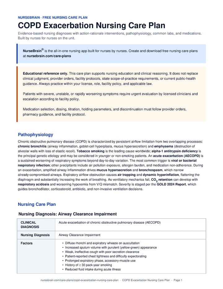
Pathophysiology
Chronic obstructive pulmonary disease (COPD) is characterized by persistent airflow limitation from two overlapping processes: chronic bronchitis (airway inflammation, goblet-cell hyperplasia, mucus hypersecretion) and emphysema (destruction of alveolar walls with loss of elastic recoil). Tobacco smoking is the leading cause worldwide; alpha-1 antitrypsin deficiency is the principal genetic etiology and may be considered in younger or non-smoking patients. An acute exacerbation (AECOPD) is a sustained worsening of respiratory symptoms beyond day-to-day variation. The most common trigger is viral or bacterial respiratory infection; other precipitants include air pollution exposure, allergen burden, and medication non-adherence. During an exacerbation, amplified airway inflammation drives mucus hypersecretion and bronchospasm, which narrow already-compromised airways. Expiratory airflow obstruction causes air trapping and dynamic hyperinflation, flattening the diaphragm and substantially increasing the work of breathing. As ventilatory mechanics fail, CO2 retention can develop with respiratory acidosis and worsening hypoxemia from V/Q mismatch. Severity is staged per the GOLD 2024 Report, which guides bronchodilator, corticosteroid, antibiotic, and non-invasive ventilation decisions.
Quick Reference
- SpO2 target: 88–92% per provider order (commonly not ≥ 92% in COPD)
- HR target: 60–100 bpm
- Antibiotic duration: 5–7 days when purulent sputum is present, per provider direction
- Systemic steroid course: Prednisone 40 mg x 5 days per GOLD 2024 and provider order
- BiPAP threshold: pH < 7.35 with rising PaCO2, per facility protocol
Common Labs
| Lab | Normal range | Significance in COPD |
|---|---|---|
| ABG – pH | 7.35–7.45 | < 7.35 with ↑ PaCO2 can reflect acute-on-chronic respiratory acidosis. Nurses report the trend and value; NIV initiation is a provider-team decision per facility protocol. |
| ABG – PaCO2 | 35–45 mmHg | Compare to patient’s known baseline when available; chronic CO2 retainers often run higher. Nurses report values outside the ordered range to the provider team. |
| ABG – PaO2 | 80–100 mmHg | PaO2 60–70 mmHg is a commonly targeted range in COPD per provider order. Higher PaO2 can worsen V/Q matching and CO2 clearance via the Haldane effect. |
| HCO3− | 22–26 mEq/L | Elevated values can reflect chronic metabolic compensation for CO2 retention. |
| WBC | 4,500–11,000/µL | Elevation can support a bacterial trigger; antibiotic decisions are provider-team decisions informed by sputum character and the Anthonisen criteria. |
| CRP | < 10 mg/L | Trend marker that can support assessment of bacterial infection and treatment response. Nurses report the trend to the provider team. |
| Sputum culture | Per facility protocol | Can help support antibiotic narrowing per provider direction. Obtain before the first antibiotic dose when feasible and ordered. |
| Chest X-ray | No acute infiltrate | Helps the provider team assess for pneumonia, pneumothorax, or pulmonary edema that may mimic AECOPD. |
| BNP | < 100 pg/mL | Can help support differentiation of AECOPD from concurrent or alternative HF exacerbation. Interpretation is a provider-team decision. |
| D-dimer | < 500 ng/mL (age-adjusted in patients > 50: age x 10 ng/mL) | PE is an under-recognized AECOPD mimic; the provider team may pursue evaluation in unexplained hypoxia. |
| Eosinophil count | < 300 cells/µL | Values > 300 can favor steroid responsiveness per GOLD 2024; corticosteroid decisions remain provider-team decisions. |
| Theophylline level | 5–15 mcg/mL | Narrow therapeutic window; toxicity concern at > 20. Nurses report values outside the ordered range and concerning symptoms. |
Common Medications
| Class | Examples | Mechanism of action | Key side effects | Nursing considerations |
|---|---|---|---|---|
| Short-acting beta-2 agonist (SABA) | Albuterol 2.5 mg (DuoNeb component) neb q4–6h scheduled; PRN q1–2h per provider order in acute exacerbation | Stimulates β2 receptors, supporting bronchial smooth-muscle relaxation and bronchodilation. | Tachycardia, tremor, hypokalemia, palpitations, anxiety. | Administer as ordered per provider direction, pharmacy guidance, and facility protocol. Check HR pre/post-neb; monitor K+ with frequent dosing per provider order; reassess wheeze and WOB after each treatment and escalate concerns per facility protocol. |
| Short-acting muscarinic antagonist (SAMA) | Ipratropium 0.5 mg (DuoNeb component) neb q4–6h scheduled; PRN q1–2h per provider order in acute exacerbation | Blocks muscarinic receptors, supporting reduction of vagal bronchoconstrictor tone. | Dry mouth, urinary retention, blurred vision (avoid in eye), paradoxical bronchospasm. | Administer as ordered. Combined with SABA as DuoNeb (albuterol 2.5 mg + ipratropium 0.5 mg) is a common first-line regimen in AECOPD per GOLD 2024 and provider order. Assess for caution flags (BPH, glaucoma) and report concerns to the provider team. |
| Inhaled corticosteroid (ICS) | Fluticasone, Budesonide | Suppresses airway inflammation; can reduce exacerbation frequency long-term. | Oral candidiasis, dysphonia, possible increased pneumonia risk, slow onset. | Administer as ordered. Teach rinse-and-spit after each dose; not used for acute rescue. Benefit may be greatest when eosinophils > 300 per GOLD 2024. |
| Systemic corticosteroid | Prednisone 40 mg PO x 5 days OR methylprednisolone IV per provider order | Reduces airway inflammation and can shorten exacerbation duration. | Hyperglycemia, hypertension, insomnia, mood lability, immunosuppression, GI bleed. | Administer as ordered. Monitor BG per facility protocol (commonly q6h) and report values outside the ordered range. No taper is typically required for the GOLD 2024 5-day course; the decision rests with the provider. Give with food when possible; teach the short-course rationale. |
| Antibiotic | Azithromycin, Doxycycline, OR Amoxicillin-clavulanate (per provider order, severity-stratified) | Bacterial coverage when sputum is purulent or mechanical ventilation is needed (Anthonisen criteria). Severity-stratified first-line options per GOLD 2024 and provider order: uncomplicated -> doxycycline or azithromycin; complicated (FEV1 < 50%, frequent exacerbator, cardiac comorbidity) -> amox-clavulanate or a respiratory fluoroquinolone; Pseudomonas risk (recent hospitalization, bronchiectasis, recent broad-spectrum antibiotics) -> antipseudomonal coverage. | GI upset, QT prolongation (azithromycin), photosensitivity (doxycycline), C. difficile. | Administer as ordered per provider direction, pharmacy guidance, and facility protocol. Commonly considered when ≥ 2 of: increased dyspnea + sputum volume + sputum purulence are present; antibiotic selection is a provider-team decision. Obtain ECG when QT-prolonging agents are ordered. |
| Methylxanthine | Theophylline, Aminophylline | Non-selective PDE inhibitor; supports bronchodilation with mild anti-inflammatory effect. | Tachyarrhythmias, seizures, nausea, tremor; narrow therapeutic index. | Administer as ordered. Target serum 5–15 mcg/mL; toxicity concern at > 20. Many drug interactions (macrolides, ciprofloxacin); rarely first-line in current practice. Report concerning levels or symptoms to the provider team. |
| Oxygen therapy | Nasal cannula 1–2 L/min; Venturi mask 24–28%, titrated per provider order | Corrects hypoxemia at the lowest effective FiO2. Excess oxygen in COPD can worsen hypercapnia, primarily via reversal of hypoxic pulmonary vasoconstriction (worsened V/Q mismatch), the Haldane effect (oxyhemoglobin releases CO2), and absorption atelectasis. | Hypercapnic respiratory failure with high FiO2, absorption atelectasis, mucosal dryness. | Administer as ordered and titrate within provider parameters. Common target SpO2 88–92% in COPD per provider order and facility protocol; titrate down when SpO2 exceeds the ordered range. Monitor for somnolence and escalate per facility protocol. |
| PDE-4 inhibitor | Roflumilast | Selective phosphodiesterase-4 inhibition; can reduce inflammation in severe COPD with chronic bronchitis. | Weight loss, diarrhea, nausea, headache; mood disturbance with new or worsening depression and suicidal ideation; contraindicated in moderate-to-severe hepatic impairment (Child-Pugh B/C). | Administer as ordered. Commonly reserved for severe COPD (FEV1 < 50%) with the chronic bronchitis phenotype and frequent exacerbations. Report mood changes to the provider team. |
References
- Makic, M. B. F., & Martinez-Kratz, M. R. (Eds.). (2023). Ackley and Ladwig’s Nursing Diagnosis Handbook: An Evidence-Based Guide to Planning Care (13th ed.). Elsevier.
- Global Initiative for Chronic Obstructive Lung Disease. (2024). Global Strategy for the Diagnosis, Management, and Prevention of COPD: 2024 Report. Retrieved from https://goldcopd.org/
- Wedzicha, J. A., Miravitlles, M., Hurst, J. R., et al. (2017). Management of COPD exacerbations: a European Respiratory Society/American Thoracic Society guideline. European Respiratory Journal, 49(3), 1600791.
Frequently Asked Questions
What is the nursing care plan for COPD?
A COPD nursing care plan organizes the assessment, nursing diagnoses, goals, interventions, and evaluation criteria for a patient with COPD Exacerbation. Diagnoses are ordered by what is currently most destabilizing for the patient.
What are the priority nursing diagnoses for COPD?
Priority diagnoses for COPD appear in the Nursing Diagnoses section above, ordered by clinical acuity. The top diagnosis should reflect what is currently most destabilizing for this specific patient.
What is the priority nursing intervention for COPD?
Priority interventions for COPD are listed in the care plan above, organized by diagnosis. The most critical actions address airway, circulation, and the highest-acuity problem first.
What complications should the nurse monitor for in COPD?
Complications to monitor for in COPD are listed within each diagnosis section above. Trend vitals, mental status, and the condition-specific red flags described in the assessment section.
A report sheet built for this patient
This is exactly the kind of patient these NurseBrain report sheets are built for. Keep everything you track on one page, all shift.