Syncope Nursing Care Plan
Syncope and presyncope nursing care plan with prioritized diagnoses, interventions, fall-risk reduction, and a printable PDF.
Nursing Care Plan
Nursing Diagnosis 1: Impaired Verbal Communication
Fall Risk related to Syncope: transient loss of consciousness from global cerebral hypoperfusion, with rapid onset, short duration, and spontaneous complete recovery as evidenced by Witnessed syncopal episode with fall; Pre-syncopal lightheadedness, diaphoresis, nausea; Orthostatic BP drop > 20/10 mmHg supine to standing; Age ≥ 65 with polypharmacy (antihypertensives, diuretics, alpha-blockers); History of recurrent unexplained syncope.
Interventions
- Obtain orthostatic vital signs (supine, sitting, standing) at 1 minute and 3 minutes per facility protocol and provider order.
- Obtain a 12-lead ECG on admission and with any recurrent prodrome per provider order; place the patient on continuous telemetry per facility protocol.
- Coordinate a comprehensive medication review with the provider team and pharmacy in patients ≥ 65 years, including a Beers-criteria screen per facility protocol.
- Document the witness account of the event: prodrome, position, duration of LOC, presence of myoclonic jerks, post-event confusion, and incontinence.
- Support provider-team risk stratification by gathering the data points needed for the San Francisco Syncope Rule (CHESS), Canadian Syncope Risk Score, or OESIL score per facility protocol.
- Assess gait, balance, and lower-extremity strength prior to first ambulation post-episode per facility protocol.
- Initiate the fall-precaution bundle per facility protocol: bed in low position, brakes locked, call light within reach, nonslip footwear, yellow armband, and door signage.
- Assist the patient to dangle legs at the bedside for 1–2 minutes before standing; require a call for first ambulation post-event per facility protocol.
- For documented orthostatic etiology, apply an abdominal binder and knee-high compression stockings (commonly 20–30 mmHg) during the day per provider order and facility protocol.
- For volume-related orthostatic or vasovagal etiology, encourage oral fluid intake when the provider has ordered liberalized fluids (commonly 2–3 L/day) and liberalize sodium per provider order when there is no HF/HTN contraindication.
- Administer prescribed midodrine or fludrocortisone as ordered; avoid midodrine within 4 hours of bedtime per provider order; recheck supine BP per facility protocol.
- Coordinate tilt-table testing per facility protocol and provider order in selected patients with unexplained recurrent syncope.
- Teach prodrome recognition: lightheadedness, sweating, nausea, blurred vision, warmth; signal to immediately sit or lie down.
- Demonstrate and have the patient return-demonstrate counter-pressure maneuvers: leg crossing with tensing, handgrip, and squatting.
- Educate on driving restrictions (state-dependent, commonly 6 months symptom-free) and to contact the provider before resuming driving.
- Teach trigger avoidance: dehydration, prolonged standing, heat, large meals, alcohol, and Valsalva (straining, coughing fits).
- Notify the provider for new arrhythmia on telemetry, chest pain, recurrent syncope, or any abnormal ECG finding.
- Coordinate cardiology consult and event monitor / Holter / implantable loop recorder for unexplained recurrent syncope per provider order and facility protocol.
Outcome: Patient experiences no syncopal episode or fall during hospitalization; Patient identifies and verbalizes plans to avoid personal triggers; Patient demonstrates at least 2 counter-pressure maneuvers.
Nursing Diagnosis 2: Impaired Cardiac Output
Cardiac Output Alteration related to Syncope: transient loss of consciousness from global cerebral hypoperfusion, with rapid onset, short duration, and spontaneous complete recovery as evidenced by Witnessed syncope with abnormal ECG (long QT, AV block, ischemic changes, WPW); New arrhythmia on telemetry; Exertional syncope or syncope without prodrome; Known structural heart disease (aortic stenosis, HOCM); Family history of sudden cardiac death.
Interventions
- Monitor cardiac rhythm continuously per facility protocol and document any new arrhythmia with corresponding patient symptoms.
- Obtain serial 12-lead ECGs on admission, with any prodrome, and per provider order.
- Auscultate heart sounds for murmurs, S3, or S4; report a systolic ejection murmur or other concerning finding to the provider team.
- Monitor serial troponin per provider order.
- Assess peripheral perfusion: capillary refill, skin temperature, color, and quality of distal pulses at intervals matched to clinical acuity and facility protocol.
- Monitor urine output per facility protocol; report UOP < 30 mL/hr for 2 consecutive hours.
- Maintain reliable IV access per facility protocol; have emergency cardiac medications and defibrillator available per unit standards.
- Administer prescribed cardiac medications (beta-blocker, antiarrhythmic) as ordered; recheck HR and BP before each dose and hold per provider parameters and facility protocol.
- Review the medication administration record for QT-prolonging agents (e.g., ondansetron, azithromycin, fluoroquinolones, haloperidol) in patients with baseline long QT; flag concerns to the provider team and pharmacy per facility protocol.
- Provide rest periods between care activities and help limit exertional triggers (Valsalva, straining) in known HOCM or aortic stenosis per provider order.
- Coordinate placement of cardiac monitors (telemetry, Holter, external event recorder, or implantable loop recorder) per provider order and facility protocol.
- Teach the patient and family the signs of recurrence: prodrome, palpitations, chest pain; to lie flat immediately and call for help.
- Educate on the importance of medication adherence and not abruptly stopping beta-blockers or antiarrhythmics without provider direction.
- For patients receiving an ICD, teach what to do if a shock fires, magnet use, driving restrictions, and infection signs at the pocket per device-team guidance.
- Escalate per facility protocol for sustained VT, complete heart block, new ST elevation, persistent SBP < 90, or recurrent syncope on telemetry.
- Coordinate cardiology and electrophysiology consults for echocardiogram, stress testing, and possible pacemaker or ICD evaluation per provider order and facility protocol.
Outcome: Patient experiences no further syncopal episodes during admission; ECG and telemetry findings are monitored and reported per facility protocol; HR and BP are monitored and reported within ordered parameters.
Nursing Diagnosis 3: Non Adherence
Knowledge Deficit related to Syncope: transient loss of consciousness from global cerebral hypoperfusion, with rapid onset, short duration, and spontaneous complete recovery as evidenced by First-time syncopal episode; Patient or family expressing fear, confusion, or misconceptions; Unfamiliarity with vasovagal triggers and counter-pressure maneuvers; Lack of understanding regarding driving restrictions; Polypharmacy with limited insight into medication contributions.
Interventions
- Assess baseline understanding of syncope, its causes, and any prior episodes using teach-back per facility protocol.
- Identify learning preferences (visual, written, verbal) and any literacy or language barriers.
- Identify the patient’s personal triggers from history: prolonged standing, heat, dehydration, pain, blood-draw, micturition.
- Provide a written discharge plan in the patient’s preferred language listing triggers, prevention, prodrome signs, and follow-up per facility protocol.
- Demonstrate counter-pressure maneuvers (leg cross + tense, handgrip, squat) and have the patient return-demonstrate each.
- Review the patient’s medication list with them, highlighting which agents the provider team and pharmacy are adjusting and why.
- Coordinate follow-up with primary care and cardiology before discharge per facility protocol; confirm the patient has appointments documented.
- Educate on prodrome recognition: lightheadedness, sweating, nausea, blurred vision, and the immediate response (sit or lie down, counter-pressure).
- Teach hydration and sodium guidance as ordered (commonly 2–3 L/day fluids and liberal salt when there is no HF/HTN contraindication) using practical measures (cups, water bottles, marked container).
- Educate on driving restrictions (state-dependent, commonly 6 months symptom-free) and when to contact the provider before resuming.
- Educate on when to call 911: chest pain, prolonged LOC, post-event confusion, palpitations, or recurrent syncope.
- Teach the patient to wear medical-alert ID identifying their syncope history, allergies, and key medications.
- Coordinate referral to fall-prevention or autonomic-clinic programs for recurrent or complex cases per facility protocol and provider order.
- Document teach-back outcomes and any persistent gaps in the chart for ambulatory follow-up per facility protocol.
Outcome: Patient verbalizes personal trigger profile; Patient demonstrates at least 2 counter-pressure maneuvers correctly; Patient describes the ordered fluid and sodium plan when there is no contraindication.
Nursing Diagnosis 4: Impaired Urinary System Function
Anxiety related to Syncope: transient loss of consciousness from global cerebral hypoperfusion, with rapid onset, short duration, and spontaneous complete recovery as evidenced by Patient verbalization of fear of recurrence or sudden death; Embarrassment about the witnessed event; Loss of driving privileges and independence; Hypervigilance toward bodily sensations (palpitations, lightheadedness); Sleep disturbance and worry.
Interventions
- Assess anxiety level using a 0–10 scale at the start of every shift and PRN per facility protocol.
- Identify the patient’s stated fears: dying, recurrence in public, loss of driving, family burden.
- Observe for physical signs: tachycardia disproportionate to clinical state, restlessness, hypervigilance to symptoms.
- Provide a calm, reassuring presence; explain workup findings in patient-friendly terms as they return.
- Explain procedures and findings before performing them.
- Cluster cares to allow uninterrupted rest periods when clinical state allows.
- Limit non-essential nighttime stimuli (overhead lights, loud conversations, unnecessary alarms) per facility protocol.
- Teach diaphragmatic breathing and grounding techniques the patient can use independently.
- Educate the patient and family on the realistic risk of recurrence based on the provider team’s established etiology.
- Teach progressive muscle relaxation as a sleep-onset aid.
- Coordinate with chaplaincy, social work, or psych services if anxiety persists or worsens.
- Notify the provider for severe or persistent anxiety unresponsive to non-pharmacologic measures.
Outcome: Patient verbalizes decreased anxiety; Patient demonstrates use of at least one coping strategy; Patient describes the realistic risk of recurrence based on the provider team’s workup.
Nursing Diagnosis 5: Fluid Imbalance
Fluid Volume Alteration related to Syncope: transient loss of consciousness from global cerebral hypoperfusion, with rapid onset, short duration, and spontaneous complete recovery as evidenced by Orthostatic BP drop > 20/10 mmHg supine to standing; Poor oral intake (< 1 L/day baseline); Use of loop or thiazide diuretics; Recent GI losses, fever, or heat exposure; Elevated BUN:Cr ratio > 20.
Interventions
- Monitor strict intake and output per facility protocol; calculate net 24-hour balance every shift.
- Reassess orthostatic vitals per facility protocol and after any ordered fluid bolus.
- Monitor BUN, creatinine, and BUN:Cr ratio per provider order.
- Assess mucous membranes, skin turgor, and capillary refill every shift.
- Encourage oral fluid intake when the provider has ordered liberalized fluids (commonly 2–3 L/day) and offer fluids hourly while awake per facility protocol, when there is no HF, ESRD, or HTN contraindication.
- Coordinate with the dietitian to liberalize sodium per provider order in appropriate patients (commonly up to 6–10 g/day per ACC/AHA 2017).
- Administer prescribed IV fluids on schedule and monitor for signs of overload (crackles, JVD, edema) per facility protocol.
- Apply abdominal binder and 20–30 mmHg knee-high compression stockings during the day per provider order and facility protocol.
- Teach the patient to drink fluids on a schedule rather than waiting for thirst.
- Educate on situations that may call for increased intake: heat, exercise, GI illness, alcohol.
- Teach recognition of dehydration: dry mouth, dark urine, dizziness on standing, fatigue.
- Notify the provider for orthostatic drop unresponsive to ordered volume, persistent UOP < 30 mL/hr, or signs of overload.
- Coordinate medication reconciliation with the provider team and pharmacy to surface diuretics, antihypertensives, vasodilators, and other volume-depleting agents per facility protocol.
Outcome: Patient meets the ordered oral intake target (commonly 2–3 L/day) when there is no HF/HTN contraindication; Orthostatic vitals are monitored and reported within ordered parameters; Urine output is monitored and reported within ordered parameters.
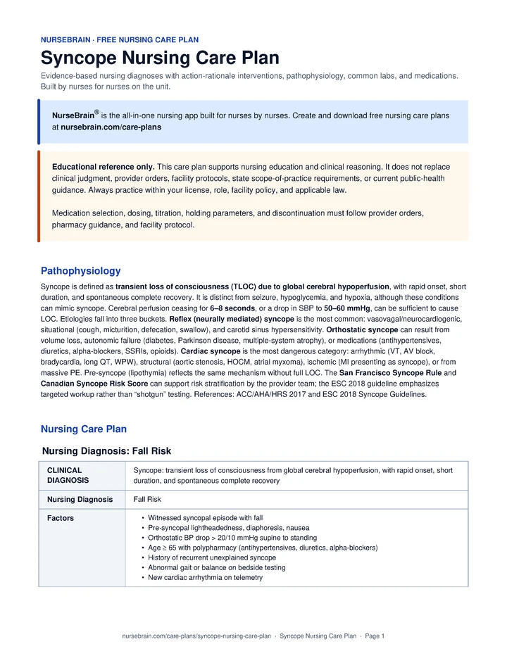
Pathophysiology
Syncope is defined as transient loss of consciousness (TLOC) due to global cerebral hypoperfusion, with rapid onset, short duration, and spontaneous complete recovery. It is distinct from seizure, hypoglycemia, and hypoxia, although these conditions can mimic syncope. Cerebral perfusion ceasing for 6–8 seconds, or a drop in SBP to 50–60 mmHg, can be sufficient to cause LOC. Etiologies fall into three buckets. Reflex (neurally mediated) syncope is the most common: vasovagal/neurocardiogenic, situational (cough, micturition, defecation, swallow), and carotid sinus hypersensitivity. Orthostatic syncope can result from volume loss, autonomic failure (diabetes, Parkinson disease, multiple-system atrophy), or medications (antihypertensives, diuretics, alpha-blockers, SSRIs, opioids). Cardiac syncope is the most dangerous category: arrhythmic (VT, AV block, bradycardia, long QT, WPW), structural (aortic stenosis, HOCM, atrial myxoma), ischemic (MI presenting as syncope), or from massive PE. Pre-syncope (lipothymia) reflects the same mechanism without full LOC. The San Francisco Syncope Rule and Canadian Syncope Risk Score can support risk stratification by the provider team; the ESC 2018 guideline emphasizes targeted workup rather than “shotgun” testing. References: ACC/AHA/HRS 2017 and ESC 2018 Syncope Guidelines.
Quick Reference
- Cardiac mortality: Up to 30% / 1 yr if untreated
- Counter-pressure: Squat, leg cross, hand grip
- Orthostatic drop: > 20 / 10 mmHg ≤ 3 min
- Beat-to-beat BP: High-risk / recurrent
- Tilt-table: Limited — unexplained recurrent VVS
Common Labs
| Lab | Normal range | Significance in Syncope |
|---|---|---|
| 12-lead ECG | Normal sinus | Commonly obtained in syncope workup to support evaluation for long QT, Brugada, WPW, AV block, and ischemia per provider order. Nurses obtain the tracing per facility protocol and report concerning findings. |
| Troponin | < 0.04 ng/mL | Supports the provider team’s assessment for MI presenting as syncope; can also be elevated in PE. Nurses trend serial values per provider order and report concerning patterns. |
| BMP | Na 135–145 / K 3.5–5.0 / Glu 70–110 | Electrolytes and glucose support the broader differential; hypoglycemia can mimic syncope. Nurses report values outside reference range to the provider team. |
| BNP / NT-proBNP | BNP < 100 pg/mL | Elevation can support a cardiac etiology or CHF contribution; interpretation is by the provider team. |
| CBC | Hgb 12–17 g/dL | Anemia can contribute to orthostatic syncope and reduced cerebral O2 delivery. Nurses report values outside reference range. |
| Pregnancy test (hCG) | Negative | Commonly obtained per facility protocol in women of childbearing age before imaging or medications, per provider order. |
| D-dimer | < 500 ng/mL | Supports PE workup when the provider team’s pre-test probability is moderate-to-high (e.g., Wells score). |
| Drug / EtOH screen | Negative | Intoxication and substance use can mimic or trigger syncope; obtain per provider order. |
| Beat-to-beat BP / autonomic testing | Stable to standing | May be considered for autonomic dysfunction workup in unexplained or recurrent cases, per provider order and facility protocol. |
| Holter / event recorder / ILR | No arrhythmia | Selection is a provider-team decision based on symptom frequency per ESC 2018: Holter (daily sx), external event recorder (weekly), ILR (monthly or less). Symptom-rhythm correlation is generally considered the gold standard. |
Common Medications
| Class | Examples | Mechanism of action | Key side effects | Nursing considerations |
|---|---|---|---|---|
| First-line (vasovagal) | Reassurance, salt + fluids, counter-pressure | Volume expansion and skeletal-muscle pump can help maintain preload during prodrome. | Generally minimal; watch for sodium overload in HF/HTN per provider order. | Reinforce as ordered. Teach prodrome recognition and counter-pressure maneuvers. When the provider has ordered liberalized fluids (commonly 2–3 L/day) and sodium, support the plan and monitor for signs of overload, per facility protocol. |
| Midodrine | ProAmatine 2.5–10 mg PO TID | Selective α1-agonist; peripheral vasoconstriction can raise BP. | Supine hypertension, piloerection, urinary retention, scalp paresthesia. | Administer as ordered per provider direction and facility protocol. Commonly not dosed within 4 hours of bedtime; nurses verify the ordered schedule, check supine BP per facility protocol, and escalate per provider hold parameters (e.g., SBP > 160). |
| Fludrocortisone | Florinef 0.1–0.2 mg PO daily | Mineralocorticoid; renal Na+/water retention supports volume expansion. | ↓ K+, ↑ BP, edema, weight gain, HF exacerbation. | Administer as ordered per provider direction. Nurses monitor K+ per ordered schedule, document daily weights, and report signs of fluid overload per facility protocol. Used cautiously in HF/HTN at the provider team’s discretion. |
| Beta-blocker | Propranolol 10–20 mg PO BID (POTS) | Can blunt β-mediated tachycardia in POTS; evidence in pure vasovagal is mixed. | Bradycardia, hypotension, fatigue, bronchospasm. | Administer as ordered. Nurses check HR and BP pre-dose and hold per provider parameters (commonly HR < 50 or SBP < 90), per facility protocol. Educate the patient not to stop the medication abruptly without provider direction. |
| SSRI | Paroxetine, Sertraline | Modulates central serotonin tone; may help reduce recurrent neurocardiogenic episodes in selected patients. | Nausea, sexual dysfunction, hyponatremia (SIADH), bleeding risk. | Administer as ordered per provider direction. Evidence is limited; selection and reassessment are provider-team decisions. Nurses monitor Na+ per ordered schedule and report concerning findings. |
| Pacemaker | Dual-chamber, rate-drop response | Paces during cardioinhibitory pauses to support patients with pause-dependent syncope. | Pocket infection, lead displacement, pneumothorax (insertion). | Coordinate referral to cardiology and electrophysiology per facility protocol when the provider team identifies pacemaker candidacy. Post-op: nurses follow facility protocol for site checks, ipsilateral arm activity restrictions (commonly 4–6 weeks), and device ID card education. |
| ICD | Single- or dual-lead implantable cardioverter-defibrillator | Detects and terminates VT/VF; can support patients at risk for sudden cardiac death. | Inappropriate shocks, infection, lead failure, psychological impact. | Coordinate referral to electrophysiology per facility protocol when the provider team identifies ICD candidacy. Teach the patient what to do if a shock fires, driving restrictions per state, and magnet-kit use as directed by the device team. |
| Medication review | Antihypertensives, diuretics, alpha-blockers, antiarrhythmics, vasodilators, opioids (elderly) | Removing or adjusting a pharmacologic contributor can be one of the most modifiable factors in orthostatic and drug-induced syncope, especially in older adults. | Rebound HTN if abrupt; loss of intended drug effect; arrhythmia if antiarrhythmic is changed. | Coordinate a full medication review (including Beers-criteria screen for adults 65+) with the provider team and pharmacy per facility protocol. Anticoagulation changes are particularly high-risk and stay with the provider team. Nurses do not independently start, stop, or adjust antihypertensives, antiarrhythmics, vasodilators, or anticoagulants. |
References
- Makic, M. B. F., & Martinez-Kratz, M. R. (Eds.). (2023). Ackley and Ladwig’s Nursing Diagnosis Handbook: An Evidence-Based Guide to Planning Care (13th ed.). Elsevier.
- Shen, W. K., Sheldon, R. S., Benditt, D. G., et al. (2017). 2017 ACC/AHA/HRS Guideline for the Evaluation and Management of Patients With Syncope. Journal of the American College of Cardiology, 70(5), e39–e110.
- Brignole, M., Moya, A., de Lange, F. J., et al. (2018). 2018 ESC Guidelines for the diagnosis and management of syncope. European Heart Journal, 39(21), 1883–1948.
Frequently Asked Questions
What is the nursing care plan for Syncope?
A Syncope nursing care plan organizes the assessment, nursing diagnoses, goals, interventions, and evaluation criteria for a patient with Syncope. Diagnoses are ordered by what is currently most destabilizing for the patient.
What are the priority nursing diagnoses for Syncope?
Priority diagnoses for Syncope appear in the Nursing Diagnoses section above, ordered by clinical acuity. The top diagnosis should reflect what is currently most destabilizing for this specific patient.
What is the priority nursing intervention for Syncope?
Priority interventions for Syncope are listed in the care plan above, organized by diagnosis. The most critical actions address airway, circulation, and the highest-acuity problem first.
What complications should the nurse monitor for in Syncope?
Complications to monitor for in Syncope are listed within each diagnosis section above. Trend vitals, mental status, and the condition-specific red flags described in the assessment section.
A report sheet built for this patient
This is exactly the kind of patient these NurseBrain report sheets are built for. Keep everything you track on one page, all shift.